. 2005 Aug;25(15):6509-20.
doi: 10.1128/MCB.25.15.6509-6520.2005.

Regulation of p53-MDMX interaction by casein kinase 1 alpha

Affiliations

Regulation of p53-MDMX interaction by casein kinase 1 alpha

Lihong Chen et al. Mol Cell Biol. 2005 Aug.

Abstract

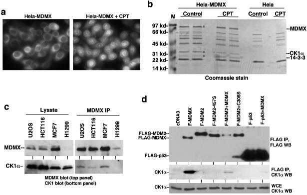
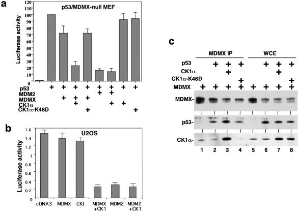
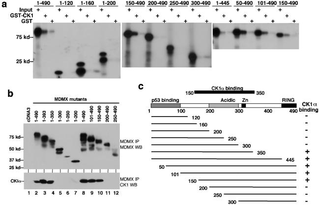
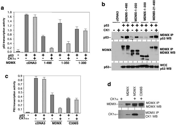
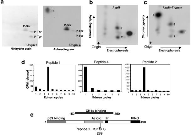
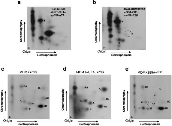
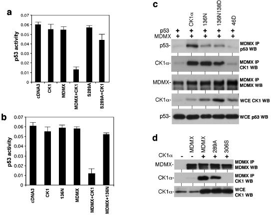
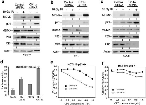
MDMX is a homolog of MDM2 that is critical for regulating p53 function during mouse development. MDMX degradation is regulated by MDM2-mediated ubiquitination. Whether there are other mechanisms of MDMX regulation is largely unknown. We found that MDMX binds to the casein kinase 1 alpha isoform (CK1alpha) and is phosphorylated by CK1alpha. Expression of CK1alpha stimulates the ability of MDMX to bind to p53 and inhibit p53 transcriptional function. Regulation of MDMX-p53 interaction requires CK1alpha binding to the central region of MDMX and phosphorylation of MDMX on serine 289. Inhibition of CK1alpha expression by isoform-specific small interfering RNA (siRNA) activates p53 and further enhances p53 activity after ionizing irradiation. CK1alpha siRNA also cooperates with DNA damage to induce apoptosis. These results suggest that CK1alpha is a functionally relevant MDMX-binding protein and plays an important role in regulating p53 activity in the absence or presence of stress.


Figures

FIG. 1.
FIG. 1.
CK1α forms a complex with MDMX. (a) HeLa cells stably transfected with FLAG-tagged human MDMX were stained using anti-FLAG M2 antibody before or after treatment with 0.5 μM camptothecin (CPT) for 16 h. (b) Coomassie blue staining of affinity-purified MDMX and associated proteins. The two marked bands were identified as CK1α and 14-3-3τ by mass spectrometric peptide sequencing. Control purification was performed using HeLa cells to exclude background. The positions of molecular mass markers (M) (in kilodaltons) are shown to the left of the gel. (c) Cell lines expressing different levels of endogenous MDMX were analyzed by MDMX IP/CK1α Western blotting. The relative expression levels of MDMX and CK1α were determined by direct Western blotting of identical amounts of whole-cell extract from each cell line. (d) H1299 cells transfected with FLAG-tagged MDMX (F-MDMX), F-MDM2, and F-p53-281G mutant were analyzed by anti-FLAG IP and anti-CK1α Western blotting (WT) to detect coprecipitation of endogenous CK1α. Cotransfection of nontagged MDMX was tested for mediating formation of trimeric complexes. F-MDM2-457S is a RING domain mutant for control. C306S is an MDMX zinc finger mutant defective for CK1α binding. WCE, whole-cell extract.
FIG. 2.
FIG. 2.
CK1α stimulates MDMX binding and inhibition of p53. (a) p53 activation of BP100-luciferase reporter was detected after transient transfection into MDMX/p53 double-null 41.4 mouse embryo fibroblasts (MEF). The inhibitory effects of MDMX and CK1α were determined by cotransfection. CK1α-K46D is a kinase-deficient mutant for control. (b) Endogenous p53 activity in U2OS cells were measured by transfection of BP100-luciferase. Inhibition by MDMX and CK1α was determined by cotransfection and luciferase assay. (c) Binding efficiency between MDMX and p53 was determined by transient cotransfection of H1299 cells with MDMX and p53 expression plasmids, followed by MDMX IP/p53 Western blotting. Whole-cell extracts (WCE) were analyzed to confirm similar levels of protein expression.
FIG. 3.
FIG. 3.
Mapping of CK1α-binding region on MDMX. (a) MDMX deletion mutants were generated by in vitro translation and incubated with glutathione agarose beads loaded with GST-CK1α. Bound MDMX proteins were detected by SDS-PAGE and autoradiography. The positions of molecular mass markers (in kilodaltons) are shown to the left of the gel. (b) MDMX deletion mutants were transiently cotransfected with CK1α expression plasmid into H1299 cells. MDMX was immunoprecipitated using 8C6 (epitope located between positions 101 to 150) or a rabbit polyclonal serum (for mutants without the 8C6 epitope). Coprecipitated CK1α was detected by Western blotting (WB). Expression and immunoprecipitation of MDMX mutants were confirmed by Western blotting (top panel). (c) Diagram of MDMX deletion mutants and summary of binding results.
FIG. 4.
FIG. 4.
CK1α binding to MDMX is required for p53 regulation. (a) p53 activation of BP100-luciferase reporter was detected after transient transfection into MDMX/p53 double-null 41.4 cells. The inhibitory effects of MDMX mutants and stimulation by CK1α were determined by cotransfection. (b) Binding efficiency between MDMX mutants and p53 was determined by transient cotransfection of H1299 cells with MDMX, p53, and CK1α expression plasmids, followed by MDMX IP/p53 Western blotting (WB). Whole-cell extracts (WCE) were analyzed to confirm similar levels of p53 expression. (c) MDMX-C306S zinc finger point mutant was not regulated by CK1α in a luciferase reporter assay similar to the results shown in panel a. (d) MDMX-C306S mutant did not bind CK1α after cotransfection into H1299 cells.
FIG. 5.
FIG. 5.
Phosphorylation of MDMX by CK1α. (a) H1299 cells were transiently transfected with MDMX and CK1α and incubated with 32Pi for 3 h. MDMX was immunoprecipitated by 8C6 and detected by autoradiography. Duplicate samples were analyzed by MDMX Western blotting (WB) to confirm expression. The positions of molecular mass markers (in kilodaltons) are shown to the left of the gel. (b) MCF7 cells were immunoprecipitated with 8C6, and the immunocomplex was incubated with [γ-32P]ATP. CKI-7 (70 μM) was used to inhibit CK1 in the reaction. Cells were treated with DNA-damaging agents (10 Gy gamma radiation or 1 μM camptothecin [CPT]) for 6 h. H1299 cells expressing very low-level MDMX were used as a control. Phosphorylation of MDMX was detected by autoradiography, and protein levels were determined by Western blotting. (c) His6-MDMX was expressed in E. coli, purified on a Ni2+-nitrilotriacetic acid column, and immunoprecipitated by 8C6. The immunocomplex was incubated with GST-CK1α and [γ-32P]ATP. Phosphorylation of MDMX was detected by autoradiography, and the multiple lower bands were confirmed to be truncated forms of GST-MDMX by Western blotting (WB).
FIG. 6.
FIG. 6.
Identification of in vitro CK1α phosphorylation site on MDMX. (a) His6-MDMX phosphorylated by GST-CK1α and [γ-32P]ATP in vitro was hydrolyzed by HCl, and phosphoamino acids were analyzed by two-dimensional electrophoresis. Ninhydrin stain indicates the positions of phosphoamino acid standards. P-Ser, phosphorylated serine. (b and c) His6-MDMX phosphorylated by GST-CK1α and [γ-32P]ATP was digested with Asp-N or Asp-N/trypsin. Phosphopeptides were analyzed by electrophoresis, followed by chromatography on a cellulose plate. (d) Phosphopeptides recovered from panels b and c were subjected to manual Edman degradation, and the release of radioactivity in each cycle was measured to determine the position of the phosphoamino acid from the N terminus. (e) Serine 289 was identified as the major CK1α phosphorylation site on the basis of Edman degradation results and sequence inspection.
FIG. 7.
FIG. 7.
Confirmation of serine 289 phosphorylation in vitro and in vivo. (a, b) Wild-type His6-MDMX or MDMX-289A mutant was phosphorylated by GST-CK1α and [γ-32P]ATP in vitro, followed by Asp-N digestion and two-dimensional peptide mapping. Mutation of serine 289 eliminated the major phosphopeptide spot 1. (c, d, e) 293T cells transfected with MDMX, MDMX and CK1α, and MDMX-289A mutant were labeled for 4 h with 32Pi. MDMX was immunoprecipitated with 8C6, digested with Asp-N, and analyzed by two-dimensional peptide mapping. Spot A migrated at the same position as the S289-containing peptide 1 in vitro, was enhanced by coexpression of CK1α, and was eliminated by the 289A mutation.
FIG. 8.
FIG. 8.
Phosphorylation of MDMX serine 289 by CK1α is required for functional regulation. (a) U2OS cells were transfected with BP100-luciferase reporter and expression plasmids for MDMX, MDMX-S289A mutants, and CK1α. Luciferase activity was determined after 24 h as an indicator of endogenous p53 activity. (b) U2OS cells were transfected with BP100-luciferase reporter and expression plasmids for MDMX, CK1α, and CK1α-D136N. The effects of the combinations on endogenous p53 activity were determined after 24 h. (c) H1299 cells were transfected with p53 and MDMX expression plasmids in combination with wild-type CK1α and CK1α with point mutations. The effects of the kinase-deficient mutants on p53-MDMX binding efficiency were determined after 48 h by MDMX IP and p53 Western blotting (WB). The ability of CK1α-D136N and CK1α-D136N-K138D mutants to bind MDMX was confirmed by MDMX IP and CK1α Western blotting. WCE, whole-cell extracts. (d) H1299 cells were transfected with MDMX mutant and CK1α expression plasmids. The ability of MDMX-289A to bind CK1α was determined by MDMX IP and CK1α Western blotting (WB). MDMX-306S served as a negative control.
FIG. 9.
FIG. 9.
Inhibition of CK1α expression by RNAi activates p53. (a, b, c) Cells were transfected with control or CK1α-specific siRNA (200 nM) for 48 h and treated with 10 Gy gamma irradiation (IR) for 6 h. MDM2 and p21 levels were determined by Western blotting. (d) U2OS cells stably transfected with p53-responsive BP100-luciferase reporter were treated as in panel c, and luciferase activity was determined using identical amount of cell lysate. Con Si, control siRNA; IR + CKI Si, gamma irradiation and CK1α-specific siRNA. (e, f) HCT116 cells were transfected with control siRNA (Con siRNA) or CK1α-specific siRNA (CK1 siRNA) (200 nM) for 24 h and treated with camptothecin (CPT) at the indicated concentrations for 24 h. Cell survival was determined by the MTT assay. OD450, optical density at 450 nm.

References

    1. Amit, S., A. Hatzubai, Y. Birman, J. S. Andersen, E. Ben-Shushan, M. Mann, Y. Ben-Neriah, and I. Alkalay. 2003. Axin-mediated CKI phosphorylation of beta-catenin at Ser 45: a molecular switch for the Wnt pathway. Genes Dev. 16:1066-1076. - PMC - PubMed
    1. Baker, S. J., S. Markowitz, E. R. Fearon, J. K. Willson, and B. Vogelstein. 1990. Suppression of human colorectal carcinoma cell growth by wild-type p53. Science 249:912-915. - PubMed
    1. Blattner, C., T. Hay, D. W. Meek, and D. P. Lane. 2002. Hypophosphorylation of Mdm2 augments p53 stability. Mol. Cell. Biol. 22:6170-6182. - PMC - PubMed
    1. Brockman, J. L., S. D. Gross, M. R. Sussman, and R. A. Anderson. 1992. Cell cycle-dependent localization of casein kinase I to mitotic spindles. Proc. Natl. Acad. Sci. USA 89:9454-9458. - PMC - PubMed
    1. Chen, J., V. Marechal, and A. J. Levine. 1993. Mapping of the p53 and mdm-2 interaction domains. Mol. Cell. Biol. 13:4107-4114. - PMC - PubMed

Publication types

MeSH terms