. 2005 Aug;25(15):6846-56.
doi: 10.1128/MCB.25.15.6846-6856.2005.

Compound genetic ablation of nidogen 1 and 2 causes basement membrane defects and perinatal lethality in mice

Affiliations

Compound genetic ablation of nidogen 1 and 2 causes basement membrane defects and perinatal lethality in mice

Bernhard L Bader et al. Mol Cell Biol. 2005 Aug.

Abstract

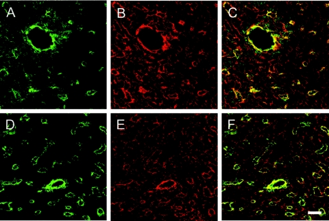
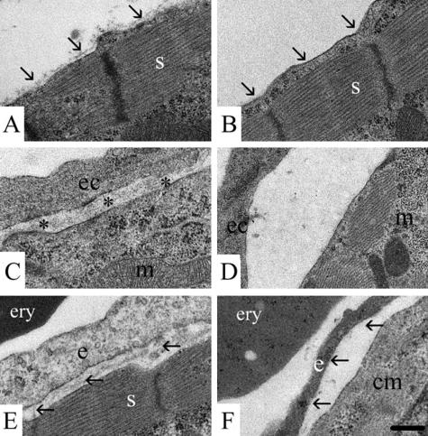
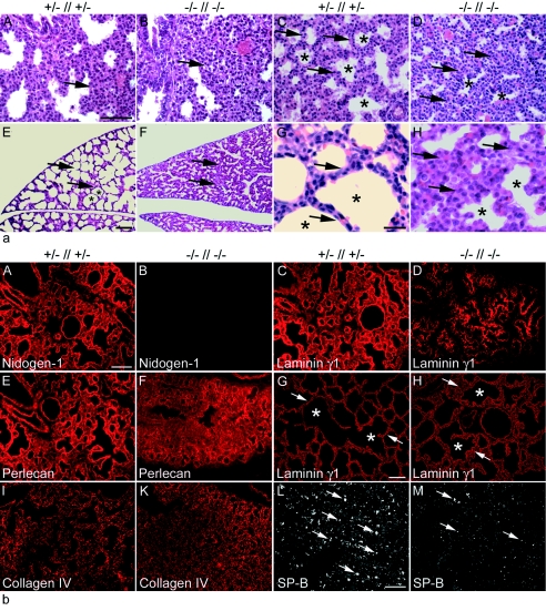
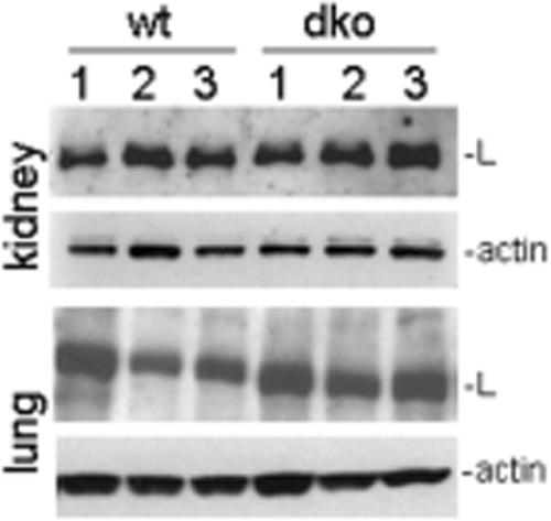
Nidogen 1 and 2 are basement membrane glycoproteins, and previous biochemical and functional studies indicate that they may play a crucial role in basement membrane assembly. While they show a divergent expression pattern in certain adult tissues, both have a similar distribution during development. Gene knockout studies in mice demonstrated that the loss of either isoform has no effect on basement membrane formation and organ development, suggesting complementary functions. Here, we show that this is indeed the case. Deficiency of both nidogens in mice resulted in perinatal lethality. Nidogen 1 and 2 do not appear to be crucial in establishing tissue architecture during organ development; instead, they are essential for late stages of lung development and for maintenance and/or integrity of cardiac tissue. These organ defects are not compatible with postnatal survival. Ultrastructural analysis suggests that the phenotypes directly result from basement membrane changes. However, despite the ubiquitous presence of nidogens in basement membranes, defects do not occur in all tissues or in all basement membranes, suggesting a varying spectrum of roles for nidogens in the basement membrane.


Figures

FIG. 1.
FIG. 1.
Histology (a) and immunofluorescence microscopy (b) of cardiac tissues from nidogen double-null embryos and neonates. (a) Hematoxylin-and-eosin-stained transverse sections of hearts from control littermates (A to D) and double-null mice (E to H) at E17.5 (A and E) and P0 (B to D and F to H) are shown. The nidogen-deficient hearts (E) were slightly smaller in size than the hearts of control embryos (A). (A, B, E, and F) Arrows, compact zones of the ventricular wall; (C, D, G, and H) asterisks, areas shown at different magnifications; arrows, cell-cell contacts. (A and E) c, endocardial cushion; l, left ventricle; r, right ventricle; ra, right atrial chamber; s, interventricular septum; t, tricuspid valve. Bars, 200 μm (A and E), 50 μm (B, C, F, and G), and 10 μm (D and H). (b) Immunofluorescence was performed on embryonic heart sections at E18.5 using rabbit antisera against nidogen 1 (A and B), nidogen 2 (C and D), laminin γ1 (E and F), perlecan (G and H), and collagen type IV (I, I*, K, K*, and L). Deposition of all basement membrane components is detectable in control sections (A, C, E, G, I, and I*), whereas in mutant cardiac tissues, the staining intensities appeared to be reduced (F, H, K, K*, and L). Areas of the cardiac muscle tissue with smaller (arrows) and larger (asterisks) blood vessels in I and K are shown at a higher magnification in I* and K*. In some sections from nidogen-deficient hearts, collagen IV staining of basement membranes of capillaries (L) appears to vary, with some showing a normal (arrow) and others showing an irregular (arrowheads) or sometimes patchy staining pattern. Bars, 50 μm (A to K, K*, I*, and L) and 25 μm (inset in L).
FIG. 2.
FIG. 2.
Expression and distribution pattern of laminin γ1 (red signal) and PECAM-1 (green signal) in cryosections from hearts of control (A to C) and nidogen double-null (D to F) embryos at E18.5. Cryosections were processed for indirect immunofluorescence and visualized by confocal microscopy. Both color channels were merged to demonstrate codistribution (yellow signal) of both immunofluorescence staining signals in the corresponding sections (C and F). Bar, 20 μm.
FIG. 3.
FIG. 3.
Electron micrographs of ultrathin sections of hearts of control littermates (A, C, E) and of nidogen double-null mutants (B, D, E) at E18.5 (A, B, E, and F) and at E16.5 (C and D). Heart cardiomyocytes with sarcomer (s) of a control embryo with the basement membrane marked by arrows (A) and of a double-null mutant where the basement membrane is lacking (B) are shown. For the endocardium, the endocardial basement membrane (asterisks) is present between the endocardial cell (ec) and the cardiomyocyte with a mitochondrium (m) in control embryos (C). In the double-null embryos, the basement membrane is missing (D). Capillaries in the heart muscle of a control embryo and a double-null mutant with an erythrocyte (ery) are shown. The basement membrane is present (arrows) underneath the endothelial cell (e) adjacent to a cardiomyocyte (cm) with a sarcomer (s) in the littermate control (E), whereas in the nidogen double-null mutant, the basement membrane is lacking (arrows). Bar, 0.25 μm.
FIG. 4.
FIG. 4.
Histological analysis of lung tissue from embryos and neonates. (a) Hematoxylin-and-eosin-stained transverse sections of lungs from comparable regions at E17.5 (A and B), E18.5 (C and D), and P0 (E to H) are shown. Development of the lung tissue from the mutant (B and D) appears delayed compared to that of control littermates (A and C). At birth, neonatal lungs from control pups show typically dilated peripheral airway saccules (E and G), whereas nidogen-deficient lungs appear immature (F and H). Arrows, mesenchyme; asterisks, saccules. Bars, 50 μm (A to F) and 10 μm (G and H). (b) Immunofluorescence of lung sections from control (A, C, E, I, and L) and nidogen double-null (B, D, F, K, and M) embryos at E18.5 and (G and H) at P0. Rabbit antisera against the basement membrane components nidogen 1 (A and B), laminin γ1 (C, D, G, H), perlecan (E and F), collagen type IV (I and K), and SP-B (L and M) were used. Prominent deposition of all basement membrane proteins is detectable in control sections (A, C, E, G, I), whereas deposition appears less pronounced at E18.5 in sections from nidogen-deficient mutants (D, F, K). However, sections from lungs of mutant animals after birth showed a laminin staining of lung basement membranes comparable to that seen in control littermates (G and H). Immunostaining for SP-B appears significantly reduced in mutant (M) compared to control (L) lungs. (G and H) Arrows and asterisks mark terminal airways. Bars, 50 μm.
FIG. 5.
FIG. 5.
Immunoblot analysis of protein extracts from kidneys and lungs isolated from three control (wild type [wt] [lanes 1 to 3]) and three nidogen double-null (dko [lanes 1 to 3]) embryos at E18.5. Twenty micrograms of total protein was separated under reducing conditions on 4 to 12% SDS-polyacrylamide gels and, after transfer, incubated with primary antibodies directed either against laminin γ1 (L) or actin, which was used as a loading control.
FIG. 6.
FIG. 6.
Analysis of kidney formation and function from nidogen-deficient embryos and neonates. Hematoxylin-and-eosin-stained sections of kidneys of control (A) and nidogen double-null (B) mice at P0 are shown. Most nidogen-deficient mice formed kidneys that were histologically normal, and immunostaining for laminin γ1 (C, control; D, nidogen double-null mutant) showed a characteristic linear pattern in the glomerulus and under most tubules. Electron microscopy of kidney at E18.5 revealed a normal glomerulus with the typical dual basement membrane being present and in tight contact with the overlying podocytes (e, glomerular epithelial cell; p, podocyte) in control littermates (E) and nidogen double-null embryos (F). However, the tubular basement membrane was often thickened, discontinuous, or even absent (arrows) in double-null kidneys (H), whereas in control kidneys (G), the tubular basement membrane is present (arrows). t, tubular epithelial cell. Bars, 200 μm (A and B), 50 μm (C and D), and 0.25 μm (E to H). Urine removed from the bladders of littermates shortly after birth was analyzed by polyacrylamide gel electrophoresis, and the gels were scanned (I, double-null mutant; K, control). There was a marginal increase in lower-molecular-weight components in the urine of nidogen double-null mice.
FIG. 7.
FIG. 7.
Most nidogen double-null animals showed little or no gross renal phenotype (A); however, a minority of nidogen-null mice had either renal dysgenesis or agenesis. Agenesis could be unilateral or bilateral (B). While the range of changes occurring in the developed kidneys included hydronephrosis, the remaining kidney in B shows an obvious dilated and tortous ureter (arrows), which could be gross (C), while other kidneys displayed polycystic defects marked by arrows (D). ad, adrenal gland; b, bladder; ep, epididymus; k, kidney; t, testis.

Similar articles

Cited by

References

    1. Ackley, B. D., S. H. Kang, J. R. Crew, C. Suh, Y. Jin, and J. M. Kramer. 2003. The basement membrane components nidogen and type XVIII collagen regulate organization of neuromuscular junctions in C. elegans. J. Neurosci. 23:3577-3587. - PMC - PubMed
    1. Arikawa-Hirasawa, E., H. Watanabe, H. Takami, J. R. Hassell, and Y. Yamada. 1999. Perlecan is essential for cartilage and cephalic development. Nat. Genet. 23:354-358. - PubMed
    1. Aumailley, M., C. Battaglia, U. Mayer, R. Nischt, R. Timpl, and J. W. Fox. 1993. Nidogen mediates the formation of ternary complexes of basement membrane components. Kidney Int. 43:7-12. - PubMed
    1. Bader, B. L., H. Rayburn, D. Crowley, and R. Hynes. 1998. Extensive vasculogenesis, angiogenesis, and organogenesis precede lethality in mice lacking αv integrins. Cell 95:507-519. - PubMed
    1. Breitkreutz, D., N. Mirancea, C. Schmidt, R. Beck, U. Werner, H.-J. Stark, M. Gerl, and N. Fusenig. 2004. Inhibition of basement membrane formation by a nidogen-binding laminin γ1-chain fragment in human skin-organotypic cocultures. J. Cell Sci. 117:2611-2622. - PubMed

Publication types

MeSH terms

LinkOut - more resources