. 2005 Aug;15(8):1061-72.
doi: 10.1101/gr.4004805. Epub 2005 Jul 15.

A functional survey of the enhancer activity of conserved non-coding sequences from vertebrate Iroquois cluster gene deserts

Affiliations

A functional survey of the enhancer activity of conserved non-coding sequences from vertebrate Iroquois cluster gene deserts

Elisa de la Calle-Mustienes et al. Genome Res. 2005 Aug.

Abstract

Recent studies of the genome architecture of vertebrates have uncovered two unforeseen aspects of its organization. First, large regions of the genome, called gene deserts, are devoid of protein-coding sequences and have no obvious biological role. Second, comparative genomics has highlighted the existence of an array of highly conserved non-coding regions (HCNRs) in all vertebrates. Most surprisingly, these structural features are strongly associated with genes that have essential functions during development. Among these, the vertebrate Iroquois (Irx) genes stand out on both fronts. Mammalian Irx genes are organized in two clusters (IrxA and IrxB) that span >1 Mb each with no other genes interspersed. Additionally, a large number of HCNRs exist within Irx clusters. We have systematically examined the enhancer activity of HCNRs from the IrxB cluster using transgenic Xenopus and zebrafish embryos. Most of these HCNRs are active in subdomains of endogenous Irx expression, and some are candidates to contain shared enhancers of neighboring genes, which could explain the evolutionary conservation of Irx clusters. Furthermore, HCNRs present in tetrapod IrxB but not in fish may be responsible for novel Irx expression domains that appeared after their divergence. Finally, we have performed a more detailed analysis on two IrxB ultraconserved non-coding regions (UCRs) duplicated in IrxA clusters in similar relative positions. These four regions share a core region highly conserved among all of them and drive expression in similar domains. However, inter-species conserved sequences surrounding the core, specific for each of these UCRs, are able to modulate their expression.


Figures
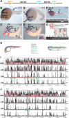
Figure 1.
Figure 1.
Expression pattern of zebrafish and Xenopus IrxB genes and distribution of highly conserved non-coding regions within the IrxB locus. (A) Intergenic distances between the three IrxB genes in human chromosome 16. (B–G) Expression pattern of zebrafish and Xenopus IrxB genes. Lateral views of 24-hpf (B, D) or 48-hpf (F) zebrafish embryos, and stage 35 Xenopus embryos (C, E). (B) Zebrafish Irx3a is expressed in neural tissues and in the pronephros. (C) Xenopus Irx3 mRNA is detected in similar domains. In addition, it is detected in the ectodermal layer of the branchial arches and in the future heart region (inset). (D) Zebrafish Irx5a is present in neural tissues. (E) Xenopus Irx5 is expressed in the brain and in the neural tube in a pattern similar to that of Irx3. In addition it is expressed in the eye and in the future heart region (inset) but not in the otic vesicle, pronephros, or head epidermis. (F) In zebrafish, Irx6 is initially expressed at 48 hpf, as determined by RT-PCR (bottom). At this stage, in whole mounts Irx6 is detected in different neural domains and in the notochord (top). (G) Xenopus Irx6 mRNA onsets of expression occurs at stage 35 (bottom), as determined by RT-PCR. Compare Irx6 initial expression with the control Histone H4 mRNA shown below. (1c) One-cell stage; (s9–50) stages 9–50. Transverse sections of a stage 40 Xenopus laevis embryo at different levels, indicated by lines in drawing at top [(a) midbrain, (b) hindbrain, (c) spinal cord], show that Irx6 is expressed in the nervous system and in the notochord. (mb) Midbrain; (hb) hindbrain; (mhb) midbrain–hindbrain boundary; (sp) spinal cord; (ov) otic vesicle; (e) ectodermal layer of the branchial; (ey) eye; (p) pronephros; (cg) cement gland; (n) notochord. (H) Color-coded schematic representation of the zebrafish and Xenopus regions with IrxB gene expression. (I, J) VISTA view of the occurrence of conserved sequence domains in the gene deserts between the Irx3 and the Irx5 (I) or Irx5 and Irx6 (J) genes from vertebrate IrxB clusters. Shown from top to bottom are mouse vs. human (M/H), chick vs. human (C/H), Xenopus tropicalis vs. human (X/H), and Fugu vs. human (F/H) global alignments. Colored peaks (purple, coding; pink, non-coding) indicate regions of at least 100 bp and 75% similarity. Gray, green, and magenta asterisks mark the amplified genomic regions with no enhancer activity, with enhancer activity in zebrafish, and with enhancer activity in Xenopus, respectively. Letters in the colored peaks are referred to in the panels shown in Figure 2 (F/H row in I), Figure 3 (X/H row in I), and Figure 4 (F/H and X/H rows in J).
Figure 2.
Figure 2.
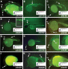
Highly conserved non-coding regions (HCNRs) located between Irx3 and Irx5 genes show enhancer activity in zebrafish. Lateral views of 24-hpf zebrafish showing enhanced green fluorescent protein localization promoted by different HCNRs (A–L). Insets show magnifications of some of the corresponding expression domains. The number in the lower righthand corner of each panel corresponds to the position of the human homologous region in chromosome 16 (NCBI build 33) as shown in Figure 1I. For better comparison, illustrations show the expression domains in which the HCNRs are active. Color codes are as those in Figure 1. Note that this is an oversimplification scheme, as many enhancers are active in subdomains of these territories. (fb) Forebrain; (mb) midbrain; (hb) hindbrain; (sp) spinal cord; (ey) eye; (n) notochord.
Figure 3.
Figure 3.
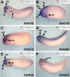
Highly conserved non-coding regions (HCNRs) located between Irx3 and Irx5 genes show enhancer activity in Xenopus. Lateral views of late neurula Xenopus embryos showing enhanced green fluorescent protein mRNA localization promoted by different HCNRs (A–F). The number in the lower righthand corner of each panel corresponds to the position of the human homologous region in chromosome 16 (NCBI build 33) as shown in Figure 1I. Illustrations show a schematic representation of the expression domains in which the HCNRs are active. (fb) Forebrain; (mb) midbrain; (hb) hindbrain; (ep) epidermis; (ey) eye; (p) pronephros.
Figure 4.
Figure 4.
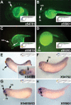
Highly conserved non-coding regions (HCNRs) located between Irx5 and Irx6 genes show enhancer activity in zebrafish and Xenopus.(A–D) Lateral views of 24-hpf zebrafish embryos showing distribution of enhanced green fluorescent protein (EGFP) promoted by different HCNRs. (E–H) Lateral views of late neurula Xenopus embryos showing EGFP mRNA localization. Inset in E shows a higher magnification of the embryo from a ventral view. The number in the lower righthand corner of each panel corresponds to the position of the human homologous region in chromosome 16 (NCBI build 33) as shown in Figure 1J. Illustrations show a schematic representation of the expression domains in which the HCNRs are active. (fb) Forebrain; (mb) midbrain; (hb) hindbrain; (ey) eye; (h) heart territory.
Figure 5.
Figure 5.
Highly conserved non-coding regions (HCNRs) are functionally active in different vertebrates. (A–C) Lateral views of 24-hpf zebrafish showing of enhanced green fluorescent protein (EGFP) fluorescence in the midbrain (mb) promoted by mouse (A), Fugu (B), or zebrafish (C) HCNR 54390. (D–E) Zebrafish 54390 placed 5′ of Xenopus Irx3 (D) or opsin (E) promoters activates EGFP expression in the same midbrain (mb) domain. Inset in D shows a higher magnification of a dorsal view of the embryo. The opsin promoter also activates EGFP expression in the eye (ey) (arrow in E). (F) Dorsal view of a stage 45 Xenopus embryo transgenic for the mouse 54390 region. EGFP expression is detected in the midbrain (mb).
Figure 6.
Figure 6.
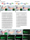
Functional comparison of Irx ultraconserved non-coding regions (UCRs). (A) Schematic representation of the vertebrate Irx clusters showing the position of the UCRs (purple boxes) between the different Irx genes. (B–E) Lateral views of late neurula Xenopus embryos showing enhanced green fluorescent protein (EGFP) mRNA localization promoted by the different Xenopus UCRs. Insets show EGFP fluorescence in stage 45 embryos transgenic for the Xenopus UCRs. Note similar expression in the brain. (mb) Midbrain; (ey) eye; (p) pronephros. (F, G) Sequence alignment of the four Xenopus UCRs (F) or Xenopus, zebrafish, and mouse UCRB1s (G). Asterisks mark identical bases. (H, I) Lateral views of stage 35 Xenopus embryos showing EGFP expression directed by zebrafish (H) or mouse (I) UCRB1s. Inset in H is a higher magnification of a living embryo transgenic for zUCRB1 showing EGFP fluorescence in the eye (arrowhead) and in the pronephros (arrow). (J) Xenopus embryo transgenic for a construct in which EGFP is directed by a 3-kb Irx3b promoter that harbors a duplicated UCRB1b. (K–O) EGFP fluorescence in Xenopus promoted by the core domain conserved between different vertebrate Irx UCRs.
Figure 7.
Figure 7.
Summary of our results. Diagram showing the distribution of the different enhancers detected in this study within the IrxB cluster. For simplicity, we have numbered these enhancers from 1–22. These numbers reflect their linear position in the genome from Irx3 to Irx6. Enhancers tested in both systems have the same number. The correspondence of these numbers with the enhancer position in human genome can be found in Supplemental Table 2. The color coding represents the different expression domains in which these enhancers are active in zebrafish (top) and Xenopus (middle). These domains are listed in the same color code. Note that this is an oversimplification scheme, as many enhancers are active in subdomains of these territories. At bottom, an illustration shows the expression domains of zebrafish and Xenopus IrxB genes.

Similar articles

Cited by

References

    1. Aparicio, S., Chapman, J., Stupka, E., Putnam, N., Chia, J.M., Dehal, P., Christoffels, A., Rash, S., Hoon, S., Smit, A., et al. 2002. Whole-genome shotgun assembly and analysis of the genome of Fugu rubripes. Science 297: 1301–1310. - PubMed
    1. Bagheri-Fam, S., Ferraz, C., Demaille, J., Scherer, G., and Pfeifer, D. 2001. Comparative genomics of the SOX9 region in human and Fugu rubripes: Conservation of short regulatory sequence elements within large intergenic regions. Genomics 78: 73–82. - PubMed
    1. Bejerano, G., Pheasant, M., Makunin, I., Stephen, S., Kent, W.J., Mattick, J.S., and Haussler, D. 2004. Ultraconserved elements in the human genome. Science 304: 1321–1325. - PubMed
    1. Bellefroid, E.J., Kobbe, A., Gruss, P., Pieler, T., Gurdon, J.B., and Papalopulu, N. 1998. Xiro3 encodes a Xenopus homolog of the Drosophila Iroquois genes and functions in neural specification. EMBO J. 17: 191–203. - PMC - PubMed
    1. Brenner, S., Elgar, G., Sandford, R., Macrae, A., Venkatesh, B., and Aparicio, S. 1993. Characterization of the pufferfish (Fugu) genome as a compact model vertebrate genome. Nature 366: 265–268. - PubMed

Publication types