. 2005 Aug;115(8):2223-33.
doi: 10.1172/JCI23755. Epub 2005 Jul 14.

The matrix component biglycan is proinflammatory and signals through Toll-like receptors 4 and 2 in macrophages

Affiliations

The matrix component biglycan is proinflammatory and signals through Toll-like receptors 4 and 2 in macrophages

Liliana Schaefer et al. J Clin Invest. 2005 Aug.

Abstract

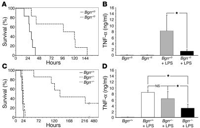
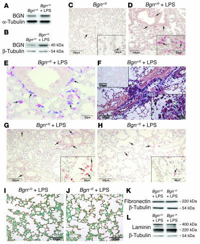
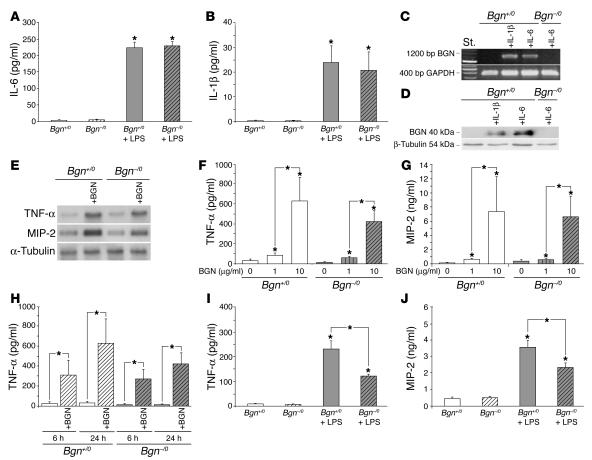
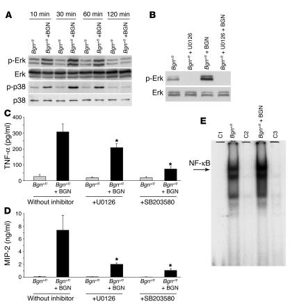
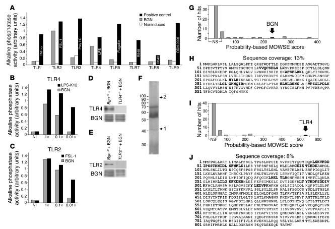
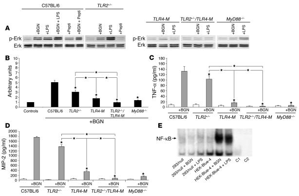
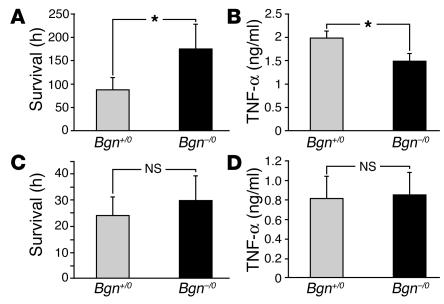
Biglycan, a small leucine-rich proteoglycan, is a ubiquitous ECM component; however, its biological role has not been elucidated in detail. Here we show that biglycan acts in macrophages as an endogenous ligand of TLR4 and TLR2, which mediate innate immunity, leading to rapid activation of p38, ERK, and NF-kappaB and thereby stimulating the expression of TNF-alpha and macrophage inflammatory protein-2 (MIP-2). In agreement, the stimulatory effects of biglycan are significantly reduced in TLR4-mutant (TLR4-M), TLR2-/-, and myeloid differentiation factor 88-/- (MyD88-/-) macrophages and completely abolished in TLR2-/-/TLR4-M macrophages. Biglycan-null mice have a considerable survival benefit in LPS- or zymosan-induced sepsis due to lower levels of circulating TNF-alpha and reduced infiltration of mononuclear cells in the lung, which cause less end-organ damage. Importantly, when stimulated by LPS-induced proinflammatory factors, macrophages themselves are able to synthesize biglycan. Thus, biglycan, upon release from the ECM or from macrophages, can boost inflammation by signaling through TLR4 and TLR2, thereby enhancing the synthesis of TNF-alpha and MIP-2. Our results provide evidence for what is, to our knowledge, a novel role of the matrix component biglycan as a signaling molecule and a crucial proinflammatory factor. These findings are potentially relevant for the development of new strategies in the treatment of sepsis.


Figures

Figure 1
Figure 1
Bgn deficiency improved survival and lowered plasma levels of TNF α in mice with LPS-induced sepsis. (A and C) Survival of male Bgn–/0 and Bgn+/0 mice (n = 6) (A) and female Bgn+/+ , Bgn+/– , and Bgn–/– mice (n = 7) (C) after a lethal dose of LPS. Two of the Bgn–/– mice survived. (B and D) Plasma levels of TNF-α in male control mice without LPS (Bgn+/0 and Bgn–/0, each group n = 3) and in Bgn+/0 (n = 5) and Bgn–/0 mice (n = 4) 90 minutes after a sublethal dose of LPS (B) and in female control Bgn+/+ mice without LPS and in Bgn+/+, Bgn+/–, and Bgn–/– mice (each group n = 3) 90 minutes after a sublethal dose of LPS (D). Data are given as means ± SD. *P < 0.05.
Figure 2
Figure 2
Increased expression of BGN in infiltrating cells of lung parenchyma in sepsis. (A and B) Northern blot of BGN mRNA normalized to α-tubulin (2 hours) (A) and Western blot of BGN core protein normalized to α-tubulin (8 hours) (B) from lungs of Bgn+/0 mice after a lethal dose of LPS versus PBS. (C and D) Immunostaining of BGN (red stain) in lungs from control (C) and septic Bgn+/0 mice (D) 8 hours after a lethal dose of LPS. (E and F) Double staining, marked by arrows, for BGN (blue) and macrophages (F4/80, red) (E) and in situ hybridization for BGN (F) in septic lungs from Bgn+/0 mice (8 hours). The lower right inset shows a magnified view of mononuclear cells expressing BGN (white arrowhead). The upper left inset represents the sense riboprobe. (G and H) Immunostaining of macrophages (F4/80, red), with numerous F4/80–positive macrophages (lower right insets), in septic lungs from Bgn+/0 (G) versus Bgn–/0 mice (H). (IL) Immunostaining of type I collagen in lungs from Bgn+/0 (brown) (I) versus Bgn–/0 mice (J) and Western blots of fibronectin (K) and laminin (L) normalized to β-tubulin in lungs from Bgn+/0 versus Bgn–/0 mice 8 hours after a lethal dose of LPS. The lower right insets in C, D, F, G, and H represent higher magnifications. Scale bars in D, H, and J apply to panels in C, G, and I, respectively.
Figure 3
Figure 3
LPS-stimulated macrophages secrete IL-6 and IL-1β, both of which induce the expression of BGN, which in turn stimulates TNF-α and MIP-2 mRNA and protein expression in macrophages. (A and B) ELISA for IL-6 (A) and IL-1β (B) in culture media from Bgn+/0 and Bgn–/0 macrophages left unstimulated or stimulated with 0.5 ng/ml LPS for 1 hour. (C and D) RT-PCR of BGN mRNA (C) and Western blot of BGN core protein (D) secreted into culture media from Bgn+/0 and Bgn–/0 macrophages 2 hours after stimulation with either IL-6 or IL-1β (both 10 ng/ml), normalized to GAPDH or β-tubulin, respectively. (E) Northern blots of TNF-α and MIP-2 mRNA (normalized to α-tubulin) in Bgn+/0 and Bgn–/0 macrophages after 6 hours of incubation with BGN (4 μg/ml). (F and G) Dose-dependent enhancement of TNF-α (F) and MIP-2 (G) concentrations in media from Bgn+/0 or Bgn–/0 macrophages cultured for 24 hours in the absence or presence of BGN (1 or 10 μg/ml). (H) Time-dependent enhancement of TNF-α concentrations in media from Bgn+/0 or Bgn–/0 macrophages cultured for 6 and 24 hours in the absence or presence of BGN (10 μg/ml). (I and J) ELISA for TNF-α (I) and MIP-2 (J) in media from Bgn+/0 or Bgn–/0 macrophages cultured for 6 hours in the absence or presence of LPS (0.5 ng/ml). Data are given as means ± SD from 3–7 animals. *P < 0.05 for macrophages with versus without BGN or LPS, respectively.
Figure 4
Figure 4
Activation of MAPKs and NF-κB by BGN in macrophages. (A) Phosphorylation of ERK and p38 in Bgn+/0 macrophages after 10–120 minutes of incubation with BGN (Western blots). (B) Effects of the MEK1/2 inhibitor (U0126) on the phosphorylation of ERK in Bgn+/0 macrophages 30 minutes after induction with BGN. (C and D) Reduced levels of TNF-α (C) and MIP-2 (D) in the presence of U0126 or SB203580 (an inhibitor of p38 MAPK) in culture media from Bgn+/0 macrophages 6 hours after induction with BGN (4 μg/ml). Data are given as means ± SD from 3–4 animals in each group. *P < 0.05 for macrophages with BGN and inhibitor versus macrophages with BGN and without inhibitor. (E) Electrophoretic mobility shift assay of nuclear extracts from Bgn+/0 macrophages cultured for 30 minutes with or without BGN. C1 to C3 represent negative controls: C1, without protein; C2 and C3, macrophages with 100 times free oligonucleotides and without (C2) or with BGN (C3).
Figure 5
Figure 5
Interaction of BGN with TLR4 and TLR2 in macrophages. (A) Screening for BGN-induced (10 μg/ml) activation of NF-κB in 293-TLR cells expressing only 1 type of TLR and alkaline phosphatase as reporter gene (gray bars). An HEK293 cell line expressing only the reporter gene was used as control (TLR). As positive control, each cell line was induced with a specific ligand (black bars). Noninduced TLR clones were used as negative controls (white bars). TLR activation is shown as activity of the secreted alkaline phosphatase in arbitrary units. (B and C) Dose-response analysis of the effect of BGN (gray bars; 1× equals 10 μg/ml) on the activation of TLR4 (B) and TLR2 (C). “NI” indicates a noninduced TLR cell line. Black bars represent positive controls (1× equals 100 ng/ml of LPS-K12 or FSL-1). (D and E) Immunoprecipitation for BGN after incubation of Bgn–/0 or TLR4–/– (D) or TLR2–/– macrophages (E) with BGN (4 μg/ml) in the presence of a cross-linker, followed by Western blot for TLR4 (D, top panel) or TLR2 (E, top panel). The bottom panels represent Western blots for BGN core protein after chondroitinase ABC treatment. (F) Colloidal Coomassie G250–stained SDS-PAGE indicating bands obtained by immunoprecipitation and analyzed by ESI/MS/MS. (GJ) ESI/MS/MS of bands labeled in F as 1 (G and H) and 2 (I and J) recognized human BGN with a probability-based score of 221 (G) and with a sequence coverage of 13% (H) and mouse TLR4 with a probability-based score of 528 (I) and with a sequence coverage of 8% (J). MOWSE, molecular weight search.
Figure 6
Figure 6
Absence of BGN-mediated stimulation of ERK, TNF-α, and MIP-2 in TLR2–/–/TLR4-M macrophages and reduced stimulation in macrophages from TLR2–/–, TLR4-M, and MyD88–/– mice. (A) Phosphorylation of ERK in C57BL/6, TLR2–/–, TLR4-M, TLR2–/–/TLR4-M, and MyD88–/– macrophages 30 minutes after incubation with BGN (4 μg/ml), LPS (2 ng/ml), or peptidoglycan (Pepti) (5 μg/ml) (Western blots). (B) Densitometric quantification of p-ERK/ERK ratio in macrophages indicated in A. Controls represent macrophages without BGN and are defined as 1 arbitrary unit. Data are given as means ± SD from 3 Western blots. (C and D) ELISA for TNF-α (C) and MIP-2 (D) in media from macrophages indicated in A after 6 hours of culture without or with BGN (4 μg/ml). Data are given as means ± SD from 7–8 animals in each group. Significant differences for TLR- or MyD88-deficient versus C57BL/6 macrophages incubated with BGN are indicated by an asterisk positioned over the respective bar: *P < 0.05. (E) BGN- or LPS-mediated (2 ng/ml) activation of NF-κB, detected after 30 minutes by EMSA in HEK-Blue-4 cells (transfected with human TLR4/MD-2/CD14 genes) versus 293/null cells. C1 (macrophages with 500 times free oligonucleotides) and C2 (without nuclear extract) represent negative controls.
Figure 7
Figure 7
Survival and plasma levels of TNF-α in zymosan- or superantigen SEB–induced sepsis. Bgn deficiency improved survival (A) and lowered plasma levels of TNF-α (B) in mice with zymosan-induced sepsis but did not affect T cell–mediated lethal shock triggered in mice by the superantigen SEB (C and D). (A) Survival of male Bgn–/0 and Bgn+/0 (n = 5) mice after a lethal dose of zymosan A. (B) Plasma levels of TNF-α in Bgn+/0 and Bgn–/0 mice (n = 5) 5 hours after a lethal dose of zymosan A. (C and D) Survival (n = 6) (C) and plasma levels of TNF-α (n = 7) (D) 90 minutes after a lethal dose of SEB simultaneously injected with D-galactosamine hydrochloride in Bgn–/0 and Bgn+/0 mice. There were no differences in the plasma levels of TNF-α between zymosan- and SEB-free mice (data not shown). *P < 0.05.

Similar articles

Cited by

References

    1. Iozzo R. The biology of the small leucine-rich proteoglycans: functional network of interactive proteins [review] J. Biol. Chem. 1999;274:18843–18846. - PubMed
    1. Ameye L, et al. Abnormal collagen fibrils in tendons of biglycan/fibromodulin-deficient mice lead to gait impairment, ectopic ossification, and osteoarthritis. FASEB J. 2002;16:673–680. - PubMed
    1. Xu T, et al. Targeted disruption of the biglycan gene leads to an osteoporosis-like phenotype in mice. Nat. Genet. 1998;20:78–82. - PubMed
    1. Moreno M, et al. Biglycan is a new extracellular component of the Chordin-BMP4 signaling pathway. EMBO J. 2005;24:1397–1405. - PMC - PubMed
    1. Weber C, et al. Biglycan is overexpressed in pancreatic cancer and induces G1-arrest in pancreatic cancer cell lines. Gastroenterology. 2001;121:657–667. - PubMed

Publication types

MeSH terms