. 2005 Jul 18:2:57.
doi: 10.1186/1743-422X-2-57.

Inhibition of Henipavirus fusion and infection by heptad-derived peptides of the Nipah virus fusion glycoprotein

Affiliations

Inhibition of Henipavirus fusion and infection by heptad-derived peptides of the Nipah virus fusion glycoprotein

Katharine N Bossart et al. Virol J. .

Abstract

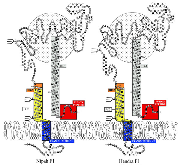
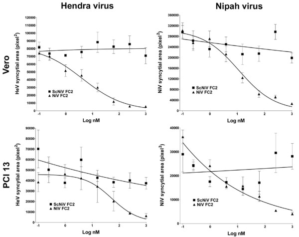
Background: The recent emergence of four new members of the paramyxovirus family has heightened the awareness of and re-energized research on new and emerging diseases. In particular, the high mortality and person to person transmission associated with the most recent Nipah virus outbreaks, as well as the very recent re-emergence of Hendra virus, has confirmed the importance of developing effective therapeutic interventions. We have previously shown that peptides corresponding to the C-terminal heptad repeat (HR-2) of the fusion envelope glycoprotein of Hendra virus and Nipah virus were potent inhibitors of both Hendra virus and Nipah virus-mediated membrane fusion using recombinant expression systems. In the current study, we have developed shorter, second generation HR-2 peptides which include a capped peptide via amidation and acetylation and two poly(ethylene glycol)-linked (PEGylated) peptides, one with the PEG moity at the C-terminus and the other at the N-terminus. Here, we have evaluated these peptides as well as the corresponding scrambled peptide controls in Nipah virus and Hendra virus-mediated membrane fusion and against infection by live virus in vitro.

Results: Unlike their predecessors, the second generation HR-2 peptides exhibited high solubility and improved synthesis yields. Importantly, both Nipah virus and Hendra virus-mediated fusion as well as live virus infection were potently inhibited by both capped and PEGylated peptides with IC50 concentrations similar to the original HR-2 peptides, whereas the scrambled modified peptides had no inhibitory effect. These data also indicate that these chemical modifications did not alter the functional properties of the peptides as inhibitors.

Conclusion: Nipah virus and Hendra virus infection in vitro can be potently blocked by specific HR-2 peptides. The improved synthesis and solubility characteristics of the second generation HR-2 peptides will facilitate peptide synthesis for pre-clinical trial application in an animal model of Henipavirus infection. The applied chemical modifications are also predicted to increase the serum half-life in vivo and should increase the chance of success in the development of an effective antiviral therapy.


Figures

Figure 1
Figure 1
Hypothetical models of the transmembrane (F1) glycoproteins of Hendra virus and Nipah virus. The models are derived by homology modeling with the known structure of the F protein of Newcastle disease virus [40]. These models are consistent protein structures predicted by the computer algorithms PHDsec [41] and TMpred [42] as described in the Methods. The heptad repeats are indicated as HR-1 (grey) and HR-2 (yellow/orange), transmembrane anchor (blue). The F2 subunit is represented by the circle behind the F1 subunit. The 36 amino acid fusion inhibitor peptide sequence used in the present study is designated as FC2 and is boxed (yellow). The equivalent location of FC2 in the HeV F1 subunit is shown for comparison.
Figure 2
Figure 2
Inhibition of Hendra virus and Nipah virus-mediated cell-cell fusion by capped C-terminal heptad peptide NiV FC2. HeLa cells were infected with vaccinia recombinants encoding HeV F and HeV G or NiV F and NiV G glycoproteins, along with a vaccinia recombinant encoding T7 RNA polymerase (effector cells). Each designated target cell type was infected with the E. coli LacZ-encoding reporter vaccinia virus vCB21R. Each target cell type (1 × 10⁵) was plated in duplicate wells of a 96-well plate. Inhibition was carried out using either capped NiV FC2 or ScNiV FC2 (control) heptad peptide. Peptides were added to the HeV or NiV glycoprotein-expressing cells (1 × 10⁵), incubated for 30 min at 37°C, and then each target cell type was added. The cell fusion assay was performed for 2.5 hr at 37°C, followed by lysis in Nonidet P-40 (1%) and β-Gal activity was quantified.
Figure 3
Figure 3
Inhibition of Hendra virus and Nipah virus-mediated cell-cell fusion by N-terminal and C-terminal (PEG10) pegylated heptad peptide NiV FC2. HeLa cells were infected with vaccinia recombinants encoding HeV F and HeV G or NiV F and NiV G glycoproteins, along with a vaccinia recombinant encoding T7 RNA polymerase (effector cells). Each designated target cell type was infected with the E. coli LacZ-encoding reporter vaccinia virus vCB21R. Each target cell type (1 × 10⁵) was plated in duplicate wells of a 96-well plate. Inhibition was carried out using either the N-terminal (N-PEG-NiV FC2) or C-terminal (C-PEG-NiV FC2) pegylated and capped heptad peptides or C-terminal pegylated scrambled control peptide (C-PEG-ScNiV FC2). Peptides were added to the HeV or NiV glycoprotein-expressing cells (1 × 10⁵), incubated for 30 min at 37°C, and then each target cell type was added The cell fusion assay was performed for 2.5 hr at 37°C, followed by lysis in Nonidet P-40 (1%) and β-Gal activity was quantified.
Figure 4
Figure 4
Immunofluorescence-based syncytia assay of Hendra virus and Nipah virus infection. Vero cells were plated into 96 well plates and grown to 90% confluence. Cells were pre-treated with heptad peptides for 30 min at 37°C prior to infection with 1.5 × 10³ TCID50/ml and 7.5 × 10² TCID50/ml of live HeV or NiV (combined with peptide). Cells were incubated for 24 hours, fixed in methanol and immunofluorescently stained for P protein prior to digital microscopy. Images were obtained using an Olympus IX71 inverted microscope coupled to an Olympus DP70 high resolution color camera and all images were obtained at an original magnification of 85×. Representative images of FITC immunofluorescence of anti-P labeled HeV and NiV syncytia are shown. A: HeV without peptide. B: HeV with C-PEG-NiV FC2. C: HeV with N-PEG-ScNiV FC2. D: NiV without peptide. E: NiV with N-PEG-NiV FC2. F: NiV with N-PEG-ScNiV FC2.
Figure 5
Figure 5
Inhibition of Hendra virus and Nipah virus infection by capped heptad peptides. Vero cells or PCI 13 cells were plated into 96 well plates and grown to 90% confluence. Cells were pre-treated with the indicated peptide for 30 min at 37°C prior to infection with 1.5 × 10³ TCID50/ml and 7.5 × 10² TCID50/ml of live HeV or NiV (combined with peptide). Cells were incubated for 24 hours, fixed in methanol and immunofluorescently labeled for P protein prior to digital microscopy and image analysis to determine the relative area of each syncytium (see Methods). The figure shows the relative syncytial area (pixel2) versus the indicated peptide concentration for HeV and NiV.
Figure 6
Figure 6
Inhibition of Hendra virus and Nipah virus infection by N-terminal and C-terminal pegylated heptad peptides. Vero cells or PCI 13 cells were plated into 96 well plates and grown to 90% confluence. Cells were pre-treated with the indicated peptide for 30 min at 37°C prior to infection with 1.5 × 10³ TCID50/ml and 7.5 × 10² TCID50/ml of live HeV or NiV (combined with peptide). Cells were incubated for 24 hours, fixed in methanol and immunofluorescently labeled for P protein prior to digital microscopy and image analysis to determine the relative area of each syncytium (see Methods). The figure shows the relative syncytial area (pixel2) versus the indicated peptide concentration for HeV and NiV.

References

    1. Murray K, Selleck P, Hooper P, Hyatt A, Gould A, Gleeson L, Westbury H, Hiley L, Selvey L, Rodwell B, et al. A morbillivirus that caused fatal disease in horses and humans. Science. 1995;268:94–97. - PubMed
    1. Chua KB, Goh KJ, Wong KT, Kamarulzaman A, Tan PS, Ksiazek TG, Zaki SR, Paul G, Lam SK, Tan CT. Fatal encephalitis due to Nipah virus among pig-farmers in Malaysia [see comments] Lancet. 1999;354:1257–1259. doi: 10.1016/S0140-6736(99)04299-3. - DOI - PubMed
    1. Barclay AJ, Paton DJ. Hendra (equine morbillivirus) Vet J. 2000;160:169–176. doi: 10.1053/tvjl.2000.0508. - DOI - PubMed
    1. Wong KT, Shieh WJ, Kumar S, Norain K, Abdullah W, Guarner J, Goldsmith CS, Chua KB, Lam SK, Tan CT, Goh KJ, Chong HT, Jusoh R, Rollin PE, Ksiazek TG, Zaki SR. Nipah virus infection: pathology and pathogenesis of an emerging paramyxoviral zoonosis. Am J Pathol. 2002;161:2153–2167. - PMC - PubMed
    1. Wang LF, Yu M, Hansson E, Pritchard LI, Shiell B, Michalski WP, Eaton BT. The exceptionally large genome of Hendra virus: support for creation of a new genus within the family Paramyxoviridae. J Virol. 2000;74:9972–9979. doi: 10.1128/JVI.74.21.9972-9979.2000. - DOI - PMC - PubMed

Publication types

LinkOut - more resources