. 2005 Jul;7(7):638-45.
doi: 10.1593/neo.04736.

Active p21-activated kinase 1 rescues MCF10A breast epithelial cells from undergoing anoikis

Affiliations

Active p21-activated kinase 1 rescues MCF10A breast epithelial cells from undergoing anoikis

Raymond E Menard et al. Neoplasia. 2005 Jul.

Abstract

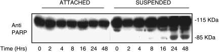
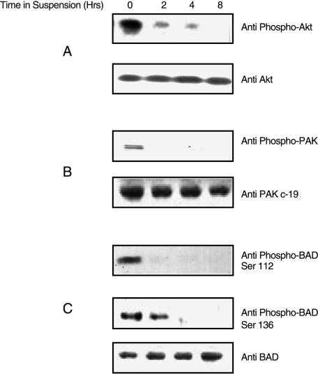
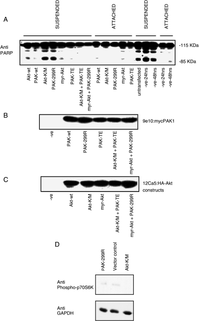
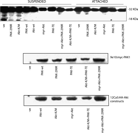
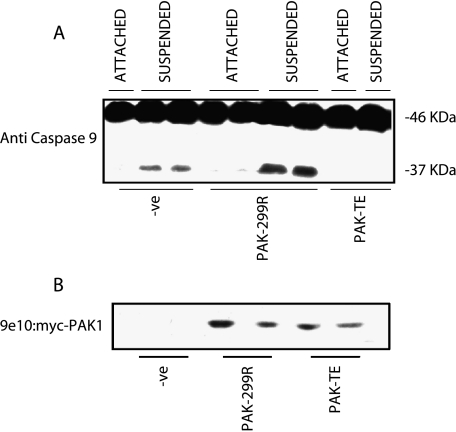
The protein kinase, PAK1, is overexpressed in human breast cancer and may contribute to malignancy through induction of proliferation and invasiveness. In this study, we examined the role of PAK1 in the survival of detached MCF10A breast epithelial cells to test whether it may also regulate the early stages of neoplasia. MCF10A cells undergo anoikis, as measured by the cleavage of caspase 3 and poly(ADP-ribose) polymerase (PARP), after more than 8 hours of detachment. Endogenous Akt, PAK1, and BAD are phosphorylated in attached MCF10A cells, but these phosphorylation events are all lost during the first 8 hours of detachment. Expression of constitutively active PAK1 or Akt suppresses the cleavage of caspase 3 and PARP in detached MCF10A cells. Co-overexpression of active PAK1 with dominant-negative Akt, or of active Akt with dominant-negative PAK1, still suppresses anoikis. Thus, Akt and PAK1 enhance survival through pathways that are at least partially independent. PAK1-dependent regulation of anoikis is likely to occur early in the apoptotic cascade as expression of dominant-negative PAK1 increased the cleavage of the upstream caspase 9, while constitutively active PAK1 inhibited caspase 9 activation. These results support a role for activated PAK1 in the suppression of anoikis in MCF10A epithelial cells.


Figures

Figure 1
Figure 1
Time course of PARP cleavage in MCF10A cells grown in suspension. Cells were grown either attached on regular culture dishes or in suspension on polyhema-coated Petri dishes for times ranging from 0 to 48 hours. PARP cleavage was detected by Western blot analysis of cell lysates for the appearance of the 85-kDa fragment.
Figure 2
Figure 2
Decrease in endogenous PAK1, Akt, and BAD phosphorylation in MCF10A cells grown in suspension. MCF10A cells were grown either attached (t = 0) or suspended for 2, 4, or 8 hours. (A) Western blots of whole cell lysates were probed with anti-phospho Akt (Ser-423) to detect active Akt, and then stripped and reprobed with anti-Akt to detect total Akt. (B) Western blots of whole cells lysates were probed with anti-phospho PAK1 (Thr-423) to detect active PAK1 (the doublet observed may be due to the fact that the antibody will also react with any endogenous PAK2 that may be present and phosphorylated on residue Thr-402), and then stripped and reprobed with anti-Pak c-19 to detect total PAK1. (C) Western blots of whole cell lysates were probed with anti-phospho-BAD (Ser-136) to detect phosphorylated BAD, then stripped and reprobed with anti-phospho-BAD (Ser-112) to detect phosphorylated BAD, and then stripped and reprobed with anti-BAD to detect total BAD.
Figure 3
Figure 3
Endogenous PAK1 is activated by EGF and LPA in MCF10A cells. MCF10A cells were treated with 100 nM EGF or 10 µM LPA for 5 minutes and lysates were prepared. The results shown are for two independent isolates of each condition that were analyzed on the same gel. After detection of active phosphorylated PAK1 (top panel), the blots were stripped and reprobed for total PAK1 (bottom panel).
Figure 4
Figure 4
Cleavage of PARP is inhibited by overexpression of active PAK1 or active Akt. MCF10A cells were transfected with plasmids encoding wild-type (wt), active (PAK-TE or myr-Akt), or dominant-negative (PAK-299R or Akt-K/M) forms of PAK1 and Akt as shown. Cells were grown for 48 hours posttransfection, counted, and split into two plates: one regular culture dish and one polyhema-coated Petri dish for a further 24 hours. Cell lysates were then processed for PARP cleavage (A), the presence of the myc tag on the transfected PAK1 constructs (B), and the presence of the HA tag on the transfected Akt constructs (C). Lysates from attached cells were also immunoblotted for phospho-p70S6K as a downstream marker of cellular Akt activity, followed by anti-GAPDH as a loading control (D).
Figure 5
Figure 5
Cleavage of caspase 3 is inhibited by overexpression of active PAK1 and active Akt. MCF10A cells were transfected with plasmids encoding wild-type (wt), active (PAK-TE or myr-Akt), or dominant-negative (PAK-299R or Akt-K/M) forms of PAK1 and Akt as shown. Cells were grown for 48 hours posttransfection, counted, and split into two plates: one regular culture dish and one polyhema-coated Petri dish for a further 24 hours. Cell lysates were then processed for detection of caspase 3 cleavage, which is revealed by the appearance of an 18-kDa fragment (upper panel). Blots were also probed with 9e10 to detect myc-tagged PAK1 constructs (middle panel) and with 12Ca5 to detect HA-Akt constructs (lower panel).
Figure 6
Figure 6
Cleavage of caspase 9 is regulated by the activation of PAK1. MCF10A cells were transfected with wild-type, active, or dominant-negative PAK1 constructs. Cells were grown for 48 hours posttransfection, counted, and split into two plates: one regular culture dish and one polyhema-coated Petri dish for a further 24 hours. Cell lysates were then processed for cleavage of caspase 9, as revealed by appearance of a 37-kDa fragment (A). Blots were also probed with 9e10 to detect myc-tagged PAK1 constructs (B).

References

    1. Frisch SM, Francis H. Disruption of epithelial cell-matrix interactions induces apoptosis. J Cell Biol. 1994;124:619–626. - PMC - PubMed
    1. Ruoslahti E, Reed JC. Anchorage dependence, integrins, apoptosis. Cell. 1994;77:477–478. - PubMed
    1. Coucouvanis E, Martin GR. Signals for death and survival: a two-step mechanism for cavitation in the vertebrate embryo. Cell. 1995;83:279–287. - PubMed
    1. Boudreau N, Sympson CJ, Werb Z, Bissell MJ. Suppression of ICE and apoptosis in mammary epithelial cells by extracellular matrix. Science. 1995;267:891–893. - PMC - PubMed
    1. Brooks PC, Montgomery AM, Rosenfeld M, Reisfeld RA, Hu T, Klier G, Cheresh DA. Integrin alpha v beta 3 antagonists promote tumor regression by inducing apoptosis of angiogenic blood vessels. Cell. 1994;79:1157–1164. - PubMed

Publication types

MeSH terms