. 2005 Sep 9;280(36):31390-6.
doi: 10.1074/jbc.M504961200. Epub 2005 Jul 15.

ATRIP oligomerization is required for ATR-dependent checkpoint signaling

Affiliations

ATRIP oligomerization is required for ATR-dependent checkpoint signaling

Heather L Ball et al. J Biol Chem. .

Abstract

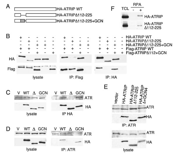
The ATM and ATR kinases signal cell cycle checkpoint responses to DNA damage. Inactive ATM is an oligomer that is disrupted to form active monomers in response to ionizing radiation. We examined whether ATR is activated by a similar mechanism. We found that the ATRIP subunit of the ATR kinase and ATR itself exist as homooligomers in cells. We did not detect regulation of ATR or ATRIP oligomerization after DNA damage. The predicted coiled-coil domain of ATRIP is essential for ATRIP oligomerization, stable ATR binding, and accumulation of ATRIP at DNA lesions. Additionally, the ATRIP coiled-coil is also required for ATRIP to support ATR-dependent checkpoint signaling to Chk1. Replacing the ATRIP coiled-coil domain with a heterologous dimerization domain restored stable binding to ATR and localization to damage-induced intranuclear foci. Thus, the ATR-ATRIP complex exists in higher order oligomeric states within cells and ATRIP oligomerization is essential for its function.


Figures

Figure 1
Figure 1
ATRIP forms oligomeric complexes. A, Myc- and Flag epitope-tagged ATRIP proteins were co-expressed in 293T cells. Proteins were immunoprecipitated using antibodies to the Myc or Flag epitopes. Isolated proteins were separated by SDS-PAGE and detected by immunoblotting with the indicated antibodies. Flag immunoprecipitations from cells expressing only Myc ATRIP was blotted with Myc antibody (middle) and Myc immunoprecipitations from cells expressing only Flag-ATRIP were blotted with Flag antibody (right) as controls for antibody specificity. B, Cells expressing near endogenous levels of HA-ATRIP were generated by retroviral infection and selection. HA immunoprecipitates from lysates of these cells or parental cells (−) were performed, separated by SDS-PAGE and blotted with ATRIP antibodies. Total cell lysates were also blotted to show the relative expression levels of the HA-ATRIP and endogenous protein.
Figure 2
Figure 2
ATR forms oligomeric complexes. A, HeLa cells were transfected with vectors encoding Flag-ATR, Myc-ATR, HA-ATRIP and/or a siRNA duplex targeted against ATRIP as indicated. Protein expression was assessed by blotting total cell lysates after SDS-PAGE separation with antibodies to the Flag, Myc or HA epitope tags. Efficiency of ATRIP siRNA knockdown was assessed by blotting with anti-ATRIP antibody. Flag or Myc immunoprecipitates were separated by SDS-PAGE and blotted with the indicated antibodies. B, Clonal cell lines stably expressing low levels of either Myc-ATR or both Myc-ATR and Flag-ATR were created by transfection and selection. Lysates from these cells or parental cells (−) were separated by SDS-PAGE and blotted with the Flag, Myc or ATR antibodies to show expression levels. Flag immunoprecipitations from the indicated cell lysates were performed, separated by SDS-PAGE and blotted with the indicated antibodies to assess the co-precipitation of Myc-ATR and ATRIP. C, A vector encoding HA-ATRIP and/or an siRNA duplex targeting ATR were transfected into HeLa cells as indicated. After two days, cells were lysed and immunoprecipitations performed with anti-HA antibodies. Immunoprecipitated proteins were separated by SDS-PAGE and blotted with the indicated antibodies. Cell lysates were also blotted to examine the levels of the proteins in the cell lysates prior to immunoprecipitation.
Figure 3
Figure 3
ATR and ATRIP oligomeric complexes are not dissociated by DNA damage. A, Vectors encoding Myc- and Flag-ATRIP proteins were transfected into HeLa cells. Cells were mock treated or treated with ionizing radiation (IR), hydroxyurea (HU), or ultraviolet light (UV) then incubated for the indicated amount of time prior to harvesting. Flag or Myc immunoprecipitations were performed from cell lysates, separated by SDS-PAGE, and blotted with Flag or Myc antibodies. B, HCT116 cells stably expressing HA-ATRIP and endogenous ATRIP at near equal levels were damaged with 50 J/m2 UV and lysed at 2 or 5 hours after damage. HA immunoprecipitates were separated by SDS-PAGE then blotted with ATRIP antibody to visualize both the HA-ATRIP and endogenous co-immunoprecipitated ATRIP proteins. Cell lysates were also blotted to ensure equal amounts of protein were analyzed. C, HEK293 cells were transfected with vectors encoding Flag-ATR, Myc-ATR, and HA-ATRIP then mock treated or exposed to 50 J/m2 of UV light. After 2 hours the cells were lysed and immunoprecipitations were performed with either anti-Myc or anti-Flag antibodies. Imunoprecipitates were separated by SDS-PAGE and blotted with Myc, Flag, or HA antibodies. D, Parental HeLa cells (−) or the HeLa cell line stably expressing low levels of Myc and Flag ATR were treated with 50 J/m2 of UV, 2mM HU or left untreated. Flag-ATR was immunoprecipitated from cell lysates and separated by SDS-PAGE. Flag-ATR and co-associated Myc-ATR and ATRIP were identified using specific antibodies as indicated.
Figure 4
Figure 4
ATRIP oligomerization is essential for stable ATRIP-ATR binding. A, Schematic diagram of wild-type ATRIP, ATRIP lacking the coiled-coil domain (ATRIPΔ112-225) and ATRIP containing a heterologous dimerization domain from the GCN4 protein (ATRIPΔ112-225+GCN4). B, ATRIP oligomerization was assessed by co-transfection of vectors encoding HA-ATRIP, HA-ATRIPΔ112-225, HA-ATRIPΔ112-225+GCN4, Flag-ATRIP, and Flag-ATRIPΔ112-225+GCN4 in HeLa cells as indicated. Flag or HA immunoprecipitates as well as cell lysate were separated by SDS-PAGE and blotted with HA or Flag antibodies. (C and D) Retroviral infection was used to generate U20S cells stably expressing full-length HA-ATRIP, HA-ATRIPΔ112-225 or HA-ATRIPΔ112-225+GCN4. Protein expression levels were examined by blotting cell lysates with either HA or ATR antibodies after SDS-PAGE separation. C, HA or D, ATR immunoprecipitates from the indicated cell lines were separated by SDS-PAGE and blotted with HA or ATR antibodies to assess protein interactions. E, U20S cells expressing HA-ATRIP, HA-ATRIPΔ112-225 or HA-ATRIPΔ112-225+GCN4 were treated with crosslinking agent before lysing cells for immunoprecipitation as in C and D above. F, HEK293 cells were transfected with vectors expressing HA-ATRIP or HA-ATRIPΔ112-225. Cells were lysed and incubated with beads bound to single stranded DNA that had either been coated with recombinant RPA (+) or mock coated (−). After extensive washing, proteins bound to the beads were denatured and separated by SDS-PAGE followed by blotting with the HA antibody. TCL is 5% of the lysate used in the pull down.
Figure 5
Figure 5
ATRIP oligomerization is required for proper ATRIP localization to damage-induced foci and ATR-dependent checkpoint signaling. A, Indirect immunoflouresence with HA antibodies was performed on U20S cells stably expressing HA-ATRIP, HA-ATRIPΔ112-225 or HA-ATRIPΔ112-225+GCN4 that had been left undamaged (−), treated with 8 Gy of IR and incubated for 3.5 hours post-damage, or treated with 2mM of HU for 5 hours. Representative micrographs of the cells are shown. B, Quantitation of HA-ATRIP, HA-ATRIPΔ112-225 and HA-ATRIPΔ112-225+GCN4 foci formation. Cells containing 5 or more easily visualized foci were counted as positive. Error bars represent standard deviations from three separate counts of 100 cells each. C, U2OS cells expressing no cDNA (vector), HA-ATRIP (WT), HA-ATRIPΔ112-225 or HA-ATRIPΔ112-225+GCN4 were transfected with ATRIP siRNA to remove endogenous ATRIP. The cDNAs were made immune to the ATRIP siRNA by mutations of wobble base pairs in the target sequence. Cells were either mock treated (−) or treated with 50 J/m2of UV light then incubated for 2 hours. Cell lysates were separate by SDS-PAGE and blotted with antibodies to HA-ATRIP, Chk1 or phospho-S345-specific antibodies to Chk1. The bar graphs are quantitations of the Chk1 phosphorylation data in which the Chk1 P-S345 signal was divided by the total Chk1 signal. All values were compared to the value obtained in the damaged HA-ATRIP (WT) expressing cells which was set at 100%.

Similar articles

Cited by

References

    1. Bakkenist CJ, Kastan MB. Cell. 2004;118:9–17. - PubMed
    1. Abraham RT. Genes Dev. 2001;15:2177–2196. - PubMed
    1. Shiloh Y. Curr Opin Genet Dev. 2001;11:71–77. - PubMed
    1. Cortez D, Guntuku S, Qin J, Elledge SJ. Science. 2001;294:1713–1716. - PubMed
    1. Brown EJ, Baltimore D. Genes Dev. 2000;14:397–402. - PMC - PubMed

Publication types

MeSH terms