. 2005 Jul 18;202(2):225-30.
doi: 10.1084/jem.20050545.

A malarial cysteine protease is necessary for Plasmodium sporozoite egress from oocysts

Affiliations

A malarial cysteine protease is necessary for Plasmodium sporozoite egress from oocysts

Ahmed S I Aly et al. J Exp Med. .

Abstract

The Plasmodium life cycle is a sequence of alternating invasive and replicative stages within the vertebrate and invertebrate hosts. How malarial parasites exit their host cells after completion of reproduction remains largely unsolved. Inhibitor studies indicated a role of Plasmodium cysteine proteases in merozoite release from host erythrocytes. To validate a vital function of malarial cysteine proteases in active parasite egress, we searched for target genes that can be analyzed functionally by reverse genetics. Herein, we describe a complete arrest of Plasmodium sporozoite egress from Anopheles midgut oocysts by targeted disruption of a stage-specific cysteine protease. Our findings show that sporozoites exit oocysts by parasite-dependent proteolysis rather than by passive oocyst rupture resulting from parasite growth. We provide genetic proof that malarial cysteine proteases are necessary for egress of invasive stages from their intracellular compartment and propose that similar cysteine protease-dependent mechanisms occur during egress from liver-stage and blood-stage schizonts.


Figures

Figure 1.
Figure 1.
A stage-specific Plasmodium papain-like cysteine protease. (A) Expression profiling of the P. berghei SERA locus. (Top) Schematic diagram of the 33.5-kb PbSERA locus. Genes conserved between all Plasmodium species are shaded gray. Rodent Plasmodium-specific genes are in white. ECP1, egress cysteine protease 1; PCP, papain-like cysteine protease with an active site cysteine; SERA, papain-like cysteine protease with an active site serine; ORF, open reading frame without homology to cysteine proteases. (Bottom) RT-PCRs from mixed blood-stage and mixed sporozoite cDNAs and genomic DNA. The merozoite- and sporozoite-specific transcripts, MSP1 and TRAP, were added as controls. (B) Oocyst-specific transcription of PbECP1. Shown is a RT-PCR analysis of PbECP1 mRNA in oocyst sporozoites (oo) and salivary gland sporozoites (sg). (C) Primary structure of Plasmodium ECP1 proteins. The putative cleavable signal sequences and the central papain-like cysteine protease domains are boxed in black and gray, respectively. Overall amino acid sequence identities of the P. yoelii and P. falciparum ECP1 orthologues (PY02063 and PFB0325c, respectively) are indicated as percentage of identical residues compared with the P. berghei sequence. (D) Conservation of the catalytic residues of the papain family within the central cysteine protease domain. The catalytic residues (in a shaded background and marked with an asterisk) are the amino-terminal cysteine and the carboxy-terminal histidine together with the asparagine, which orients the histidine imidazole ring. The glutamine (bold and marked with ‘o’) in proximity to the catalytic cysteine assists in formation of the oxyanion hole. Strictly conserved amino acid residues are boxed in gray.
Figure 2.
Figure 2.
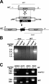
Targeted gene disruption of P. berghei ECP1. (A) Insertion strategy to generate the ecp1(-) parasites. The WT ECP1 genomic locus (WT) is targeted with an EcoRV (E)-linearized integration plasmid (pINT) containing 5′ and 3′ truncations of the ECP1 open reading frame and the dhfr/ts positive selectable marker. Upon a single crossover event, the region of homology is duplicated, resulting in two truncated, nonexpressed ecp1 copies in the recombinant locus (INT). Integration-specific test and WT primer combinations are indicated by arrows and expected fragments as lines. (B) Integration-specific PCR analysis. The successful integration event is verified by a primer combination (test) that can amplify only a signal from the INT locus. Absence of the WT signal from ecp1(-) parasites confirms the purity of the clonal population. (C) Absence of ECP1 transcripts in ecp1(-) parasites. cDNA from WT and ecp1(-) oocyst sporozoites were amplified in the presence (+) or absence (-) of reverse transcriptase (RT). Note that expression of the adjacent genes, SERA2 and ORF2 (Fig. 1 A), are not affected in the ecp1(-) parasites.
Figure 3.
Figure 3.
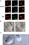
ecp1(-) oocysts generate viable sporozoites. (A) ecp1(-) oocyst sporozoites display normal gliding locomotion. Shown are representative immunofluorescence stainings of ecp1(-) and WT oocyst sporozoites with anti-PbCSP antibodies (28) and anti-TRAP antisera (29). Gliding sporozoites deposit CSP in their trails. Bars, 10 μm. (B) ecp1(-) sporozoites are confined within oocysts. Shown are interference contrast micrographs of WT and ecp1(-) oocysts. WT sporozoites are arranged radially and will eventually exit the oocyst. In contrast, ecp1(-) sporozoites do not escape the oocysts and orient in circles under continuous gliding locomotion (Video 1). Bars, 10 μm. (C) Sporozoite clusters from ecp1(-)-infected mosquitoes. Shown are micrographs from isolated and ground midguts. Free sporozoite clusters can be detected in the dissection medium indicating that lack of ECP1 alters the normal oocyst rupturing process. Bars, 10 μm.
Figure 4.
Figure 4.
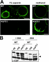
Oocysts are protected in ecp1(-) parasites. (A) ecp1(-) oocysts are resistant to permeabilization by detergents (1% saponin). The inner oocyst membrane is stained with highly diluted anti-PbCSP antibody (1:1,000). Proper CSP localization is shown in methanol-fixed oocysts. Bars, 10 μm. (B) Western blot analysis of CSP in isolated salivary gland (sg) or midgut (mg) sporozoites from WT or midgut sporozoites from ecp1(-) parasites. In addition to the typical CSP doublet (marked with asterisks), an intermediate midgut-specific band can be detected in the ecp1(-) mutant and in WT sporozoites that were isolated in the presence of 100 μM cysteine protease inhibitor E64.

References

    1. Sibley, L.D. 2004. Intracellular parasite invasion strategies. Science. 304:248–253. - PubMed
    1. Blackman, M.J. 2000. Proteases involved in erythrocyte invasion by the malaria parasite: function and potential as chemotherapeutic targets. Curr. Drug Targets. 1:59–83. - PubMed
    1. Hadley, T., M. Aikawa, and L.H. Miller. 1983. Plasmodium knowlesi: studies on invasion of rhesus erythrocytes by merozoites in the presence of protease inhibitors. Exp. Parasitol. 55:306–311. - PubMed
    1. Lyon, J.A., and J.D. Haynes. 1986. Plasmodium falciparum antigens synthesized by schizonts and stabilized at the merozoite surface when schizonts mature in the presence of protease inhibitors. J. Immunol. 136:2245–2251. - PubMed
    1. Salmon, B.L., A. Oksman, and D.E. Goldberg. 2001. Malaria parasite exit from the host erythrocyte: a two-step process requiring extraerythrocytic proteolysis. Proc. Natl. Acad. Sci. USA. 98:271–276. - PMC - PubMed

Publication types

MeSH terms