. 2005 Jul 18;202(2):239-48.
doi: 10.1084/jem.20050413.

Expansion and long-range differentiation of the NKT cell lineage in mice expressing CD1d exclusively on cortical thymocytes

Affiliations

Expansion and long-range differentiation of the NKT cell lineage in mice expressing CD1d exclusively on cortical thymocytes

Datsen G Wei et al. J Exp Med. .

Abstract

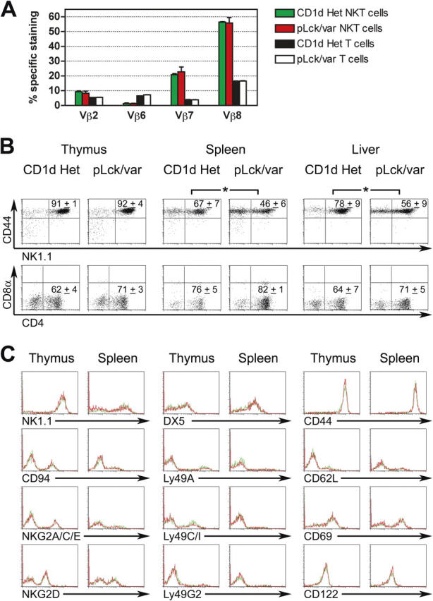
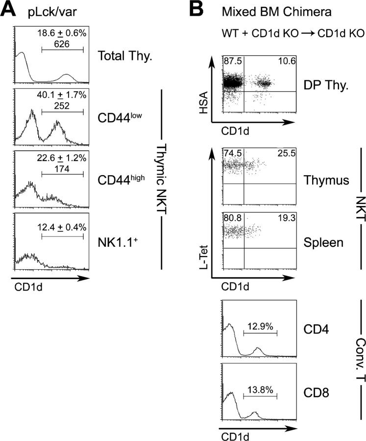
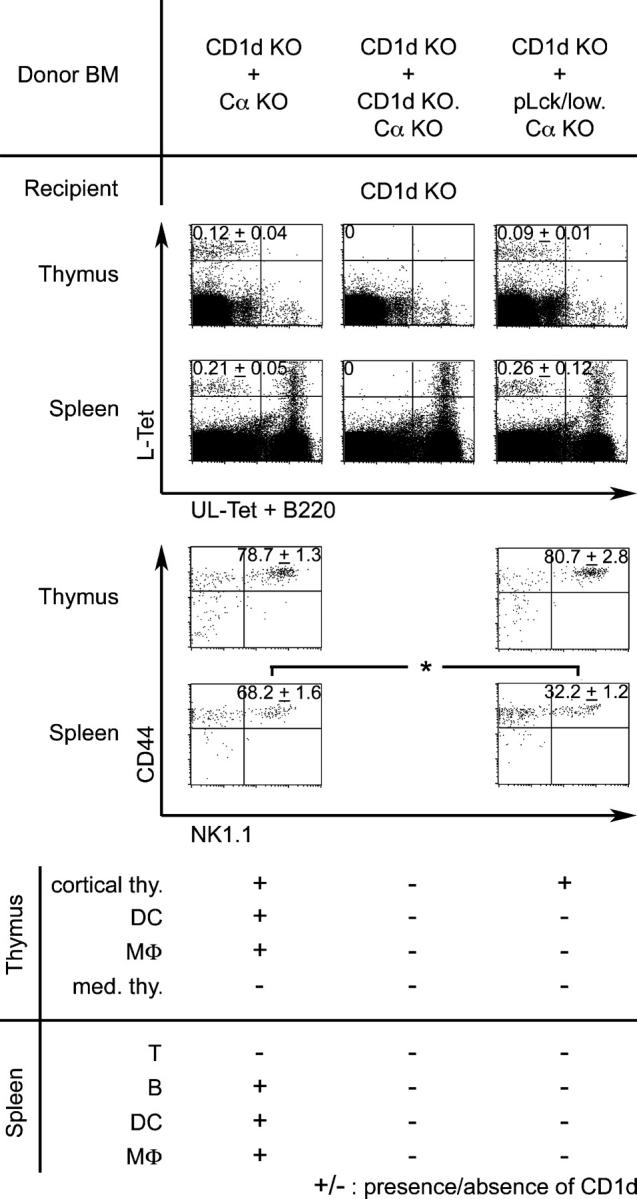
Unlike conventional major histocompatibility complex-restricted T cells, Valpha14-Jalpha18 NKT cell lineage precursors engage in cognate interactions with CD 1 d-expressing bone marrow-derived cells that are both necessary and sufficient for their thymic selection and differentiation, but the nature and sequence of these interactions remain partially understood. After positive selection mediated by CD1d-expressing cortical thymocytes, the mature NKT cell lineage undergoes a series of changes suggesting antigen priming by a professional antigen-presenting cell, including extensive cell division, acquisition of a memory phenotype, the ability to produce interleukin-4 and interferon-gamma, and the expression of a panoply of NK receptors. By using a combined transgenic and chimeric approach to restrict CD1d expression to cortical thymocytes and to prevent expression on other hematopoietic cell types such as dendritic cells, macrophages, or B cells, we found that, to a large extent, expansion and differentiation events could be imparted by a single-cognate interaction with CD1d-expressing cortical thymocytes. These surprising findings suggest that, unlike thymic epithelial cells, cortical thymocytes can provide unexpected, cell type-specific signals leading to lineage expansion and NKT cell differentiation.


Figures

Figure 1.
Figure 1.
CD1d expression and NKT cells in pLck-CD1d mice. (A) Transgenic construct for expression of CD1d cDNA under the control of the proximal Lck promoter. CD1d cDNA was inserted via the EcoRI site located in the second exon of the β-globin expression cassette. Black boxes, exons; thin lines, introns; gray box, promoter. The construct was excised via SpeI and XhoI sites from the bacterial vector, purified, and injected into fertilized eggs. (B) CD1d (red) versus I-Ab (green) expression in frozen thymic sections of three different lines of pLck-CD1d mice, pLck/low, pLck/high, and pLck/var, compared with CD1d Het (CD1d + / ) and CD1d KO (CD1d / ) littermate controls. Upper four rows are mice in the NODxC57BL/6 F1 background; bottom two rows are in the C57BL/6 background. A thin white line delineates the boundary between cortex (c) and medulla (m). Note the variegated pattern of CD1d expression in pLck/var. (C) Staining of total thymocytes and splenocytes with CD1d tetramers loaded with α-GalCer (L-Tet, y-axis) versus anti-B220 + unloaded CD1d tetramers (UL-Tet + B220, x-axis). Percentages of NKT cells ± SD, compiled from five experiments and five to eight mice per line, are shown in upper left quadrants.
Figure 2.
Figure 2.
Pattern of CD1d expression in pLck/var mice. CD1d expression on gated thymic (left column) and peripheral (right column) subsets shown in green for WT, red for pLck/var, and black for CD1d KO. Percentages and mean fluorescence intensity of CD1d+ cells are indicated above fluorescence peaks. Results are representative of three independent experiments, and more than three mice are analyzed for each cell type. (left) DN, CD48 DN and CD3ɛ; DP, CD4+8+. CD4 and CD8 are mature HSAlow single-positive cells. NKT are NK1.1+CD3ɛ+ cells. Thymic dendritic cells (DC) were enriched with anti-CD11c magnetic microbeads and further gated on CD11c+I-Ab+ for analysis. Thymic macrophages (MΦ) were enriched and gated similarly with CD11b; thymic epithelial cells (TEC) were enriched by depleting hematopoietic cells with anti-CD45 magnetic microbeads, and gating on CD45-I-Ab+ cells. (right) BM, CD3ɛ BM cells. All other subsets are gated from splenic preparations, except PMN, Gr-1+CD3ɛ polymorphonuclears from peripheral blood.
Figure 3.
Figure 3.
NKT cell phenotype in pLck/var mice. (A) TCR Vβ repertoire analysis on gated CD3ɛ+CD1d-α-GalCer tetramer+ NKT cells and mainstream CD3ɛ+ T cells in the spleen of pLck/var and CD1d Het littermate controls, as indicated. Results shown as mean + SD of data collected from nine mice in two independent experiments. (B) CD44/NK1.1 and CD4/CD8α staining of NKT cells in thymus, spleen, and liver of pLck/var mice and CD1d Het controls. Percentages and SD from 23 mice in seven experiments are indicated. *, P < 0.05 by unpaired Student's t test. (C) Analysis of NK and activation/memory markers expressed by gated CD1d-α-GalCer tetramer+ NKT cells of pLck/var (red) versus littermate CD1d Het control (green). Results representative of six pairs of pLck/var transgenic and WT littermates. Similar results were found for pLck/low mice (data not depicted).
Figure 4.
Figure 4.
BrdU incorporation by NKT cell subsets in vivo. 4 h after i.v. injection of BrdU, pLck/var and control CD1d Het showed similar frequency of BrdU+ cells among CD1d-α-GalCer tetramer+ NKT cell subsets. Similar results were found in three different experiments. Percentages of BrdU+ cells are shown in top right quadrants.
Figure 5.
Figure 5.
CD1d expression autonomously enhances NKT cell development. (A) Frequency of CD1d expression is higher among NKT cells of pLck/var mice than among total thymocytes. The difference is particularly visible at the CD44low NKT stage before extinction of the pLck promoter. Frequencies are indicated as mean ± SD of three mice. (B) In mixed WT + CD1d KO BM radiation chimeras, a higher proportion of NKT cells are CD1d-positive than expected from analysis of the DP and conventional mature CD4 and CD8 T cells. Data are representative of three individual chimeras.
Figure 6.
Figure 6.
NKT cell development in CD1d KO + pLck/low.Cα KO mixed BM chimeras. Irradiated (900 rad) C57BL/6.CD1d KO recipients of a mixture of BM cells, as indicated, were analyzed for frequency and phenotype of CD1d-α-GalCer tetramer+ NKT cells 6–8 wk after reconstitution. In these experiments, the pLck/low.Cα KO BM cells were obtained from a H-2b donor. Percentages ± SD are calculated from four individual chimeras. *, statistically significant differences (P < 0.05, unpaired Student's t test). The bottom table recapitulates the thymic and peripheral cell types expressing (+) or not expressing (−) CD1d in these chimeras.
Figure 7.
Figure 7.
Cytokine secretion by NKT cells in vivo and in vitro. (A) Mice of indicated genotypes were injected i.v. with 5 × 10⁵ α-GalCer-pulsed DCs (αGC-DC), and IFN-γ was measured in sera taken at indicated time points. Results are shown as mean ± SD of two independent experiments, each including four mice per group. (B) Early IL-4 released in vitro by cultured ∼1-mm3 splenic fragments, 90 min after i.v. injection of 1.25 μg anti-CD3ɛ in vivo. (C) Titrated numbers of sorted CD1d-α-GalCer tetramer+NK1.1+ cells were stimulated with 105 α-GalCer-pulsed DCs, before collection of supernatant for cytokine assays 48 h later. Results pooled from at least three independent experiments. Error bars represent 1 SD (not all error bars are visible). (D) CD44/NK1.1 staining of splenic CD1d-α-GalCer tetramer+ NKT cells in age-matched (6 wk) mice of indicated genotypes. Percentages and SD from three mice per group in three independent experiments are indicated. *, statistical significance by unpaired Student's t test.

Similar articles

Cited by

References

    1. Godfrey, D.I., K.J. Hammond, L.D. Poulton, M.J. Smyth, and A.G. Baxter. 2000. NKT cells: facts, functions and fallacies. Immunol. Today. 21:573–583. - PubMed
    1. Brigl, M., and M.B. Brenner. 2004. CD1: antigen presentation and T cell function. Annu. Rev. Immunol. 22:817–890. - PubMed
    1. Park, S.H., and A. Bendelac. 2000. CD1-restricted T-cell responses and microbial infection. Nature. 406:788–792. - PubMed
    1. Benlagha, K., T. Kyin, A. Beavis, L. Teyton, and A. Bendelac. 2002. A thymic precursor to the NKT cell lineage. Science. 296:553–555. - PubMed
    1. Pellicci, D.G., K.J. Hammond, A.P. Uldrich, A.G. Baxter, M.J. Smyth, and D.I. Godfrey. 2002. A natural killer T (NKT) cell developmental pathway involving a thymus-dependent NK1.1(-)CD4(+) CD1d-dependent precursor stage. J. Exp. Med. 195:835–844. - PMC - PubMed

MeSH terms