

Magnetocardiography predicts coronary artery disease in patients with acute chest pain
- PMID: 16029382
- PMCID: PMC6932599
- DOI: 10.1111/j.1542-474X.2005.00634.x
Magnetocardiography predicts coronary artery disease in patients with acute chest pain
Abstract
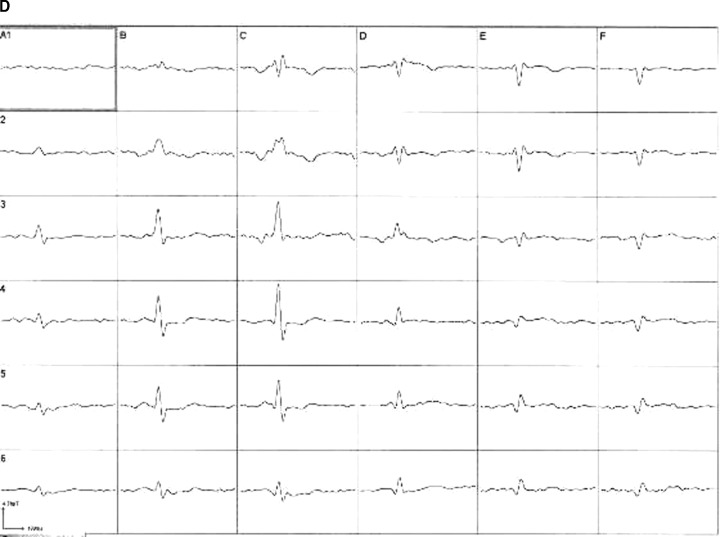
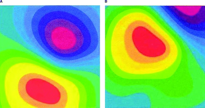
Background: The value of magnetocardiography (MCG) for the detection of cardiac electrical disturbances associated with myocardial ischemia was studied.
Methods: Sensitivity and predictivity of admission MCG for the presence of coronary artery disease (CAD) were prospectively evaluated in 264 consecutive patients presenting with acute chest pain and without ST-segment elevation. MCG findings were compared with 12-lead ECG, echocardiography (ECHO), and troponin-I in a head-to-head design. Coronary angiography was used for CAD diagnosis.
Results: The visual assessment of magnetocardiograms by the experienced reader (R1) was superior to that by the unexperienced reader (R2) and superior to the automated computer analysis. Specificity and positive predictive value of MCG by R1 were comparable with those of ECG and troponin-I (>90%), while ECHO specificity and ECHO positive predictive value were lower (76.2% and 87.9%, respectively). Sensitivity and negative predictive value of MCG were twice as high as those in the ECG, troponin-I, and ECHO tests.
Conclusion: For the prediction of CAD in patients presenting with acute chest pain and without ST-segment elevation, an admission MCG test was superior to an admission ECG, ECHO, and troponin-I. The results of the study, however, are applicable only to a highly selected population comprising patients in whom immediate coronary angiography can be performed based on their clinical course in the hospital.
Figures
References
-
- Gibler BE. Diagnosis of acute coronary syndromes in the emergency department In: Topol E. (ed.): Acute Coronary Syndromes. New York , Marcel Dekker, 2000, pp. 193–231.
-
- Gibler BE, Lewis LM, Erb RE, et al Early detection of acute myocardial infarction in patients presenting with chest pain and nondiagnostic ECGs: Serial CK‐MB sampling in emergency department. Ann Emerg Med 1990;9: 1359–1366. - PubMed
-
- Bertrand ME, Simoons ML, Fox KAA, et al Management of acute coronary syndromes: Acute coronary syndromes without persistent ST‐segment elevation. Eur Heart J 2000;21: 1–32. - PubMed
-
- Sternickel K, Tralshawala N, Bakharev A, et al Unshielded measurements of cardiac electric activity using magneto‐cardiography. Int J Bioelectromagnetism 2002;4: 189–195.
-
- Scanlon PJ, Faxon DP, Audet AM, et al ACC/AHA guidelines for coronary angiography. J Am Coll Cardiol 1999;33: 1756–1824. - PubMed
Publication types
MeSH terms
LinkOut - more resources
Full Text Sources
Other Literature Sources
Medical
Miscellaneous
