Comparative Study
. 2005 Jul 26;102(30):10676-81.
doi: 10.1073/pnas.0504922102. Epub 2005 Jul 19.

Mutually dependent secretion of proteins required for mycobacterial virulence

Affiliations
Comparative Study

Mutually dependent secretion of proteins required for mycobacterial virulence

S M Fortune et al. Proc Natl Acad Sci U S A. .

Abstract

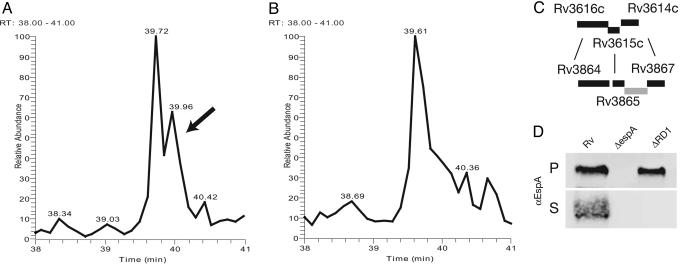
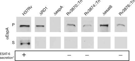
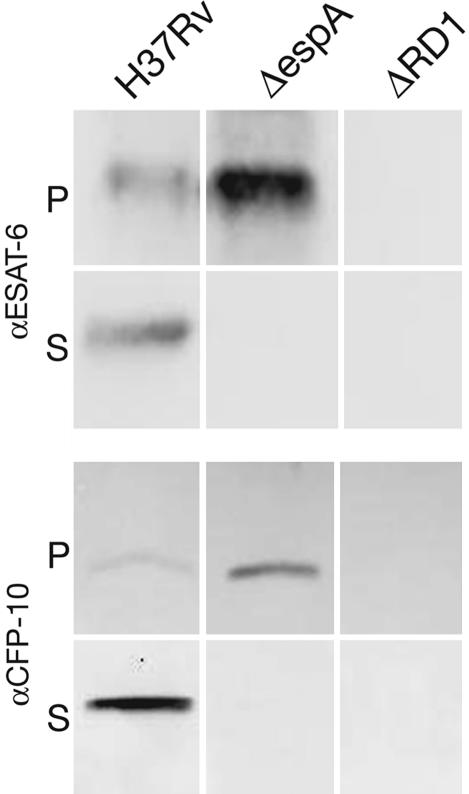
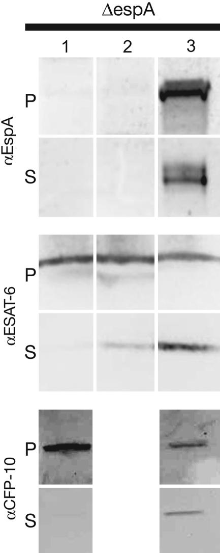
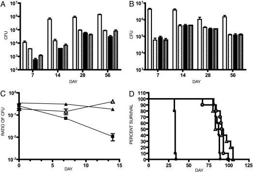
The ESX-1 locus is a region critical for full virulence in Mycobacterium tuberculosis, which encodes two secreted proteins as well as other genes involved in their secretion. The mechanism of secretion of the two proteins, ESAT-6 and CFP-10, and their function remain unknown. Using proteomic methods to search for additional proteins secreted by the ESX-1 locus, we discovered that a protein encoded by a chromosomally unlinked gene, espA, is also secreted by strains that contain the ESX-1 locus but not by strains with ESX-1 deletions. Mutations in individual ESX-1 genes, including those that encode ESAT-6 and CFP-10, were found to block EspA secretion. Surprisingly, mutants that lack espA reciprocally failed to secrete ESAT-6 and CFP-10 and were as attenuated as ESX-1 mutants in virulence assays. The results indicate that secretion of these proteins, which are each critical for virulence of pathogenic mycobacteria, is mutually dependent. The results further suggest that discerning the nature of the interaction and the structure of macromolecular complexes will provide insights into both an alternative mechanism of protein secretion and mycobacterial virulence.


Figures

Fig. 1.
Fig. 1.
EspA is present in H37Rv culture filtrates but not in RvDRD1 culture filtrates. (A and B) EspA identified by MS as present in H37Rv culture filtrates but not in RvΔRD1 culture filtrates. Culture filtrate proteins from H37Rv (A) and H37RvΔRD1 (B) were tryptically digested. In parallel runs, the resultant peptide fragments were resolved through reverse-phase chromatography and analyzed by LCQ-MS/MS. The tracing indicated by the arrow was identified as peptide KYSEGAAAGTEDAERAPVEADAGGGQK from EspA, which was one of four unique peptides identified from EspA. (C) The espA locus shares significant homology to genes upstream of the ESX-1 locus. Rv3616c, here renamed EspA, shares 31.75% identity in a 400-aa overlap with Rv3864. Rv3615c shares 36.25% identity in a 102-aa overlap with Rv3865. Rv3614c shares 54.9% identity in a 173-aa overlap with Rv3867. (D) Presence of EspA in the pellet (P) and supernatant (S) of H37Rv, H37RvΔespA (ΔespA), and H37RvΔRD1 (ΔRD1). Results are representative of more than three experiments.
Fig. 2.
Fig. 2.
Localization of EspA in wild-type M. tuberculosis and strains carrying deletions of RD1 (ΔRD1), of esat-6 (Δesat-6), and transposon insertions in the indicated genes. Cell pellet (P) and culture supernatant (S) proteins were resolved through SDS/PAGE. EspA was identified by immunoblotting with protein-specific antibody. Results are representative of at least two experiments. *, ESAT-6 secretion as published in ref. .
Fig. 3.
Fig. 3.
Localization of ESAT-6 and CFP-10 in the presence or absence of espA. Cell pellet (P) and culture supernatant (S) proteins from H37Rv, H37RvΔRD1 (ΔRD1), and H37RvΔespA (ΔespA) were resolved through SDS/PAGE. ESAT-6 and CFP-10 were identified by immunoblotting with protein-specific antibody. Results are representative of more than three experiments.
Fig. 4.
Fig. 4.
Complementation of H37RvΔespA for ESAT-6 and CFP-10 secretion. H37RvΔespA was complemented by integration of a single copy of a control vector (lane 1), epitope-tagged espA under a synthetic MOP promoter (pALE2) (lane 2) or epitope-tagged espA under its native promoter (pALE4) (lane 3) into the chromosomal attB site. Cell pellet (P) and culture supernatant (S) proteins from the complemented strain or a strain carrying the backbone vector were resolved through SDS/PAGE. EspA, ESAT-6, and CFP-10 were identified by immunoblotting with protein-specific antibody. Pellet-associated EspA expressed from pALE2 could be visualized only by overexposing the blot. Results are representative of two experiments.
Fig. 5.
Fig. 5.
Deletion of espA attenuates M. tuberculosis to a similar extent as deletion of RD1. Lungs (A) and spleen (B) colony-forming units from C57BL/6 mice infected with the indicated mycobacterial strains. The infecting inocula were determined by plating serial dilutions as follows: open bars, H37Rv, 1.9 × 10⁵; dotted bars, H37RvΔRD1, 1.6 × 10⁵; filled bars, H37RvΔespA, 1.2 × 10⁵; hashed bars, H37RvΔRD1ΔespA, 2.1 × 10⁵. Numbers represent the mean and standard deviation of four mice per time point. Each of the mutants was significantly attenuated compared to H37Rv in both organs at all time points except in the spleen colony-forming units at 28 days (P ≤ 0.01). In addition, the lung burden of RvΔRD1 was slightly but significantly higher than that of H37RvΔespA and H37RvΔRD1ΔespA at 7 days and H37Rv ΔespA at 14 days (P < 0.05). (C) C57BL/6 mice were infected with two strains of M. tuberculosis in competition. Mice were infected with a 1:3 mixture of kanamycin marked to unmarked strains as indicated: ▪, H37RvΔRD1:H37Rv; ▴, H37RvΔRD1:H37RvΔespA; ▵, H37RvΔRD1ΔespA:H37RvΔespA. The organ burden of each strain was determined at the indicated times by plating in the presence and absence of kanamycin. Numbers represent the mean and standard deviation of three mice per time point. H37RvΔRD1 was significantly attenuated relative to H37Rv over time as determined by linear regression analysis (P < 0.05). In the other two competitions, the ratios of the strains did not change significantly over time. (D) SCID mice were infected with a goal inoculum of 300 organisms. The infecting inocula, determined by plating serial dilutions, were as follows: ▪, H37Rv, 3 × 10²; ▵, H37RvΔRD1, 7 × 10²; ▾, H37RvΔespA, 23 × 102; ○, H37RvΔRD1ΔespA, 7 × 10². Ten mice were infected with each strain. The difference in survival between wild-type and mutant strains was highly statistically significant (P < 0.0001), and the times to death of RvΔespA and RvΔRD1ΔespA were not significantly different from that of RvΔRD1.

References

    1. Finlay, B. B. & Falkow, S. (1997) Microbiol. Mol. Biol. Rev. 61, 136-169. - PMC - PubMed
    1. Galan, J. E. & Collmer, A. (1999) Science 284, 1322-1328. - PubMed
    1. Cascales, E. & Christie, P. J. (2003) Nat. Rev. Microbiol. 1, 137-149. - PMC - PubMed
    1. Mahairas, G. G., Sabo, P. J., Hickey, M. J., Singh, D. C. & Stover, C. K. (1996) J. Bacteriol. 178, 1274-1282. - PMC - PubMed
    1. Pym, A. S., Brodin, P., Brosch, R., Huerre, M. & Cole, S. T. (2002) Mol. Microbiol. 46, 709-717. - PubMed

Publication types

Substances

Associated data

LinkOut - more resources