Comparative Study
. 2005 Jul 26;102(30):10460-5.
doi: 10.1073/pnas.0504249102. Epub 2005 Jul 19.

Stimulation of mammalian translation initiation factor eIF4A activity by a small molecule inhibitor of eukaryotic translation

Affiliations
Comparative Study

Stimulation of mammalian translation initiation factor eIF4A activity by a small molecule inhibitor of eukaryotic translation

Marie-Eve Bordeleau et al. Proc Natl Acad Sci U S A. .

Abstract

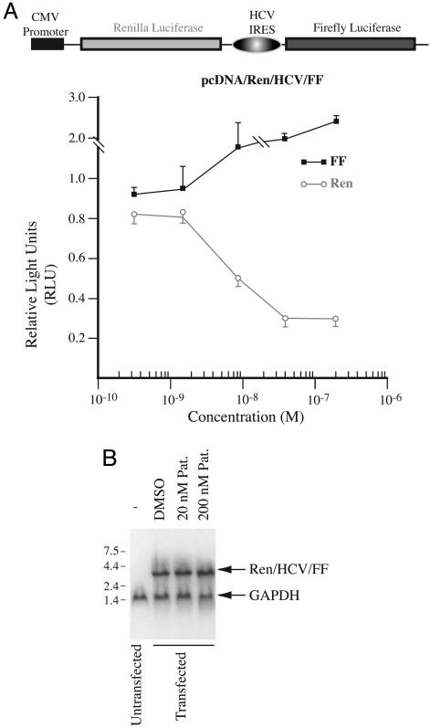
RNA helicases are the largest group of enzymes in eukaryotic RNA metabolism. The DEXD/H-box putative RNA helicases form the helicase superfamily II, whose members are defined by seven highly conserved amino acid motifs, making specific targeting of selected members a challenging pharmacological problem. The translation initiation factor eIF4A is the prototypical DEAD-box RNA helicase that works in conjunction with eIF4B and eIF4H and as a subunit of eIF4F to prepare the mRNA template for ribosome binding, possibly by unwinding the secondary structure proximal to the 5' m7GpppN cap structure. We report the identification and characterization of a small molecule inhibitor of eukaryotic translation initiation that acts in an unusual manner by stimulating eIF4A-associated activities. Our results suggest that proper control of eIF4A helicase activity is necessary for efficient ribosome binding and demonstrate the feasibility of selectively targeting DEAD-box RNA helicases with small molecules.


Figures

Fig. 1.
Fig. 1.
Inhibition of eukaryotic translation by pateamine. (A) Chemical structure of pateamine. (B) Schematic diagram of bicistronic constructs used to assess translation inhibition by pateamine. (C) Titration of pateamine in Krebs extracts programmed with bicistronic mRNAs. Translations were performed in the presence of the indicated amounts of pateamine and at a final mRNA and K+ concentration of 5 μg/ml and 100 mM, respectively. Firefly and renilla luciferase activity (RLU) were measured on a Berthold Technologies (Bad Wildbad, Germany) Lumat LB 9507 luminometer. Control translation reactions contained equivalent amounts of DMSO (data not shown). (Upper) Representative autoradiograph from an experiment performed with [35S]methionine. After separation of protein products on 10% polyacrylamide/SDS gels, the gels were treated with EN3Hance (PerkinElmer) dried, and exposed to X-Omat (Kodak) film. (Lower) Graphical representation of the effects of pateamine on translation of bicistronic mRNAs in Krebs extracts. The obtained luciferase activities were normalized to the activity obtained in the absence of compound (which was set at one). Each data point represents the average of three to seven translations, and the standard error of the mean is presented.
Fig. 2.
Fig. 2.
Pateamine inhibits ribosome recruitment. (A and B) 32P-labeled CAT and HCV/Ren mRNAs were incubated in rabbit reticulocyte lysate in the presence of 1 mM GMP-PNP or 1 mM GMP-PNP and 10 μM pateamine. After centrifugation (SW40; 39,000 rpm/3.5 h), fractions from each sucrose gradient were collected by using a Brandel (Bethesda, MD) Tube Piercer connected to an ISCO fraction collector and were individually counted. Total counts recovered from each gradient and the percent mRNA bound in 48S complexes were: (A) CAT mRNA/GMP-PNP (64,430 cpm, 21% binding) and CAT mRNA/GMP-PNP+pateamine (62,713 cpm, <0.7% binding), and (B) HCV/Ren mRNA/GMP-PNP (57,844 cpm, 7% binding) and HCV/Ren mRNA/GMP-PNP+pateamine (56,113 cpm, 6% binding). (C) eIF4A is specifically retained on a pateamine-Sepharose affinity matrix. Pateamine was coupled to epoxy-activated Sepharose, and total HL-60 cell extracts were loaded onto pateamine or control resin, washed with 0.1% Triton X-100 in 10 mM PBS, and eluted in SDS/PAGE sample buffer. Proteins were fractionated on a 4-12% polyacrylamide/SDS gel and stained with colloidal Coomassie blue. (D) Immunoblots of HL-60 extract (LOAD) and eluents from control- and pateamine-affinity resins. The antibodies used in the immunoblots are indicated. Recombinant eIF4AI, eIF4AII, and eIF4AIII proteins were used to assess antibody specificity. Recombinant eIF4AII has a higher molecular mass than recombinant eIF4AI due to additional vector-derived sequences.
Fig. 3.
Fig. 3.
Pateamine stimulates eIF4AI activity. (A) Pateamine stimulates eIF4AI-mediated ATP hydrolysis. ATPase assays were performed for the indicated times by using 1 μM γ-[32P]ATP/1 μM poly(U)/3.6 μg of recombinant eIF4AI and monitored by thin-layer chromatography. Quantitations were performed by using Fujix BAS2000 with a Fuji imaging screen, and the data are from a total of three experiments. (B) Pateamine stimulates ATP binding to eIF4AI in the presence of RNA. Crosslinking of ATP was performed with 1 μg of recombinant eIF4AI and 2.5 μCi (Ci = 37 GBq) of α-[32P]ATP (3,000 Ci/mmol) by using UV light and resolved by SDS/PAGE. The presence or absence of 7.5 μM poly(U) is indicated below. The gel was dried and exposed to x-ray film (Kodak) at -80°C for 12 h with an intensifying screen. (C) Pateamine stimulates RNA-binding activity of eIF4AI. 32P-cap-labeled CAT mRNA (105 cpm) was incubated with recombinant eIF4AI (1 μg) for 10 min at 30°C, chemically crosslinked, treated with RNase A, and resolved by SDS/PAGE. Gels were dried and exposed to x-ray film (Kodak) at -80°C for 1 h with an intensifying screen. (D) Chemical crosslinking of eIF4F to 32P-cap labeled oxidized mRNA. The presence of 10 μM pateamine is indicated at the top. Components of the eIF4F complex are labeled to the right. The gel was dried and exposed to x-ray film (Kodak) at -80°C with an intensifying screen. (E) UV light-induced crosslinking of α-[32P]ATP to eIF4Ac. Crosslinking of ATP was performed with 0.74 μg of eIF4F and 2.5 μCi of α-[32P]ATP (3,000 Ci/mmol) by using UV light and resolved by SDS/PAGE. The gel was dried and exposed to x-ray film (Kodak) at -80°C.
Fig. 4.
Fig. 4.
Pateamine stimulates eIF4AI helicase activity. (A) Reactions were performed with RNA duplexes RNA-1/RNA-11 or RNA-1/RNA-12 for 15 min at 35°C in the presence of 0.36 μg of recombinant protein. The presence or absence of 1 mM ATP or 10μM pateamine is indicated. Reactions were resolved on 12% native gels, which were dried, and exposed to x-ray film (Kodak) at -80°C for 12 h with an intensifying screen. The position of migration of duplex or radioactive single-strand RNA is indicated to the left. (B) Helicase assays performed with recombinant Ded1p protein in the presence of pateamine. Conditions were similar to those described for eIF4AI. (C) In vitro splicing reactions in the presence of pateamine. In vitro splicing reactions were performed with the AdML pre-mRNA and analyzed, as described (17). Reaction products were separated on a 15% polyacrylamide/8 M urea gel, which was dried, and exposed to X-Omat (Kodak) x-ray film at -80°C for 1 h. The position of migration of the pre-mRNA (lane 1) and spliced mRNA (lane 2) is indicated to the right. Splicing reactions were performed without pateamine (lane 3), in the presence of increasing concentrations of pateamine [lane 4 (0.5 μM), lane 5, (2 μM), and lane 6 (10 μM)], or in the absence of exogenously added ATP (lane 7).
Fig. 5.
Fig. 5.
Pateamine inhibits cap-dependent protein synthesis in vivo. (A) Dose-response experiment of pateamine on cells transfected with pcDNA/Ren/HCV/FF. HeLa cells were incubated with the indicated concentrations of pateamine for 10 h, at which time luciferase activity was measured in cell extracts. The relative luciferase activity was determined by comparing to the activity obtained in control cells exposed to DMSO. The average of two experiments is presented with the error of the mean. (B) Northern blot of RNA isolated from cells transfected with pcDNA/Ren/HCV/FF. After transfection, cells were incubated with the indicated concentrations of pateamine. RNA was isolated, fractionated on a 1.2% agarose/formaldehyde gel, and transferred to Hybond N+ (Amersham Pharmacia Biosciences). The blot was then probed with radiolabeled cDNA fragments to Ren/HCV/FF and GAPDH (position of migration indicated). Probes were produced with the Readiprime kit using the manufacturer's recommendations (Amersham Pharmacia).

References

    1. Hershey, J. W. B. & Merrick, W. C. (2000) Initiation of Protein Synthesis (Cold Spring Habor Lab. Press, Plainview, NY).
    1. Wilson, J. E., Pestova, T. V., Hellen, C. U. & Sarnow, P. (2000) Cell 102, 511-520. - PubMed
    1. Grifo, J. A., Abramson, R. D., Satler, C. A. & Merrick, W. C. (1984) J. Biol. Chem. 259, 8648-8654. - PubMed
    1. Rogers, G. W., Jr., Richter, N. J. & Merrick, W. C. (1999) J. Biol. Chem. 274, 12236-12244. - PubMed
    1. Richter-Cook, N. J., Dever, T. E., Hensold, J. O. & Merrick, W. C. (1998) J. Biol. Chem. 273, 7579-7587. - PubMed

Publication types

LinkOut - more resources