Comparative Study
. 2005 Aug;187(15):5376-86.
doi: 10.1128/JB.187.15.5376-5386.2005.

NanA, a neuraminidase from Streptococcus pneumoniae, shows high levels of sequence diversity, at least in part through recombination with Streptococcus oralis

Affiliations
Comparative Study

NanA, a neuraminidase from Streptococcus pneumoniae, shows high levels of sequence diversity, at least in part through recombination with Streptococcus oralis

Samantha J King et al. J Bacteriol. 2005 Aug.

Abstract

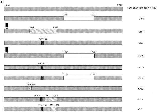
Streptococcus pneumoniae, an important human pathogen, contains at least two genes, nanA and nanB, that express sialidase activity. NanA is a virulence determinant of pneumococci which is important in animal models of colonization and middle ear infections. The gene encoding NanA was detected in all 106 pneumococcal strains screened that represented 59 restriction profiles. Sequencing confirmed a high level of diversity, up to 17.2% at the nucleotide level and 14.8% at the amino acid level. NanA diversity is due to a number of mechanisms including insertions, point mutations, and recombination generating mosaic genes. The level of nucleotide divergence for each recombinant block is greater than 30% and much higher than the 20% identified within mosaic pbp genes, suggesting that a high selective pressure exists for these alterations. These data indicate that at least one of the four recombinant blocks identified originated from a Streptococcus oralis isolate, demonstrating for the first time that protein virulence determinants of pneumococci have, as identified previously for genes encoding penicillin binding proteins, evolved by recombination with oral streptococci. No amino acid alterations were identified within the aspartic boxes or predicted active site, suggesting that sequence variation may be important in evading the adaptive immune response. Furthermore, this suggests that nanA is an important target of the immune system in the interaction between the pneumococcus and host.


Figures

FIG. 1.
FIG. 1.
Nucleotide (A) and amino acid (B) alignment to demonstrate diversity within the 3′ region of nanA from R36A, Pn13, and TIGR4. Numbering of the sequence starts from the first putative ATG within the R36A published sequence (X72967 [6]). Identical nucleotides and amino acids are illustrated by a dot, while absent sites are represented by a dash. The location of block D is boldfaced. The three 60-bp (20-amino-acid) direct tandem repeats are underlined, and a vertical line indicates the start of each repeat. A 7-bp sequence present at the start of each repeat and immediately distal to the repeats (A) is double underlined. The 15-bp (5-amino-acid) duplication within the Pn13 and TIGR4 sequence is italicized. The asterisk indicates that the protein terminates at that residue (B).
FIG. 2.
FIG. 2.
Diversity within the 5′ region of nanA. Nucleotide (A) and amino acid alignment (B) and schematic (C) of the 5′ published sequences from R36A, TIGR4, and 12 other pneumococci selected to represent a range of 5′ RFLP profiles. The numbering refers to the position within the published sequence starting at the first putative ATG within the R36A published sequence (X72967 [6]). To allow reference to the location within the published sequence, residues within the 48-bp illegitimate insertion are not numbered. Only residues displaying diversity are presented. Absent sites are shown by dashes, while dots represent identical residues (A and B). Blocks A, B, and C are indicated by underlining, italics, and boldface, respectively (A and B). The location of the recombinant blocks within the 5′ schematic is represented by diagonally striped boxes (block A), diamond-patterned boxes (block B), and white boxes (block C) (C). The 48-bp insertion is shown in black (C).
FIG. 2.
FIG. 2.
Diversity within the 5′ region of nanA. Nucleotide (A) and amino acid alignment (B) and schematic (C) of the 5′ published sequences from R36A, TIGR4, and 12 other pneumococci selected to represent a range of 5′ RFLP profiles. The numbering refers to the position within the published sequence starting at the first putative ATG within the R36A published sequence (X72967 [6]). To allow reference to the location within the published sequence, residues within the 48-bp illegitimate insertion are not numbered. Only residues displaying diversity are presented. Absent sites are shown by dashes, while dots represent identical residues (A and B). Blocks A, B, and C are indicated by underlining, italics, and boldface, respectively (A and B). The location of the recombinant blocks within the 5′ schematic is represented by diagonally striped boxes (block A), diamond-patterned boxes (block B), and white boxes (block C) (C). The 48-bp insertion is shown in black (C).
FIG. 3.
FIG. 3.
Schematic of diversity identified within the 3′ region of nanA. The numbering starts at the corresponding location within the coding sequence of the published sequence starting at the first putative ATG within the R36A published sequence (X72967 [6]). Block D is represented by diagonally striped boxes, and the 60-bp repeat sequences are represented by dark gray boxes, while the 15-bp repeat sequences are represented by light gray boxes.
FIG. 4.
FIG. 4.
Southern blot of block D and the 60-bp repeats to isolates of other streptococcal species. The repeat sequences and block D from R36A were DIG labeled and used as a probe. Five micrograms of chromosomal DNA from each strain was restricted with EcoRV, electrophoresed, and transferred to a membrane, and the Southern blot assay was performed. Isolates included were Cr7 (nanAΔR) (lane 1), Cr48 (nanAR4) (lane 2), three S. oralis isolates (lanes 4, 5, and 6), one S. mitis isolate (lane 7), three S. sanguinis isolates (lanes 8, 9, and 10), one S. cristatus isolate (lane 11), one S. constellatus subsp. constellatus isolate (lane 12), and one S. ratti isolate (lane 13). Numbers at right are molecular sizes in base pairs.

Similar articles

Cited by

References

    1. Beighton, D., and S. Alum. 1997. Use of repetitive extragenic palindromic PCR (REP-PCR) to study Streptococcus oralis. J. Dent. Res. 76:1026.
    1. Beighton, D., and R. Whiley. 1990. Sialidase activity of the “Streptococcus milleri group” and other viridans group streptococci. J. Clin. Microbiol. 28:1431-1433. - PMC - PubMed
    1. Berry, A. M., R. A. Lock, and J. C. Paton. 1996. Cloning and characterization of nanB, a second Streptococcus pneumoniae neuraminidase gene, and purification of the NanB enzyme from recombinant Escherichia coli. J. Bacteriol. 178:4854-4860. - PMC - PubMed
    1. Berry, A. M., and J. C. Paton. 2000. Additive attenuation of virulence of Streptococcus pneumoniae by mutation of the genes encoding pneumolysin and other putative pneumococcal virulence proteins. Infect. Immun. 68:133-140. - PMC - PubMed
    1. Brooks-Walter, A., D. E. Briles, and S. K. Hollingshead. 1999. The pspC gene of Streptococcus pneumoniae encodes a polymorphic protein, PspC, which elicits cross-reactive antibodies to PspA and provides immunity to pneumococcal bacteremia. Infect. Immun. 67:6533-6542. - PMC - PubMed

Publication types

Associated data