. 2005 Oct;16(10):4531-42.
doi: 10.1091/mbc.e05-04-0330. Epub 2005 Jul 19.

Myosin 2 is a key Rho kinase target necessary for the local concentration of E-cadherin at cell-cell contacts

Affiliations

Myosin 2 is a key Rho kinase target necessary for the local concentration of E-cadherin at cell-cell contacts

Annette M Shewan et al. Mol Biol Cell. 2005 Oct.

Abstract

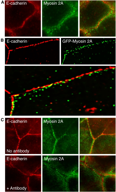
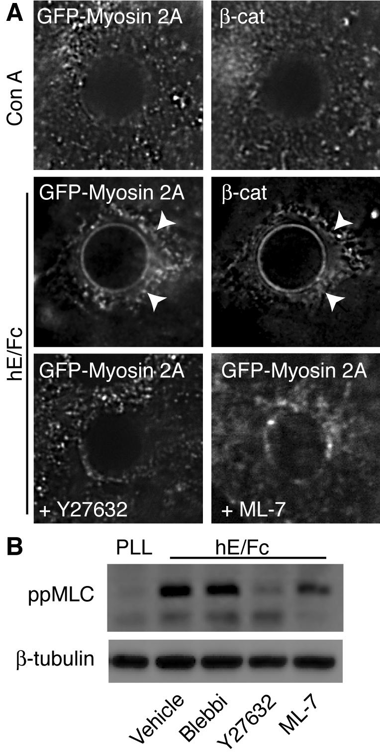
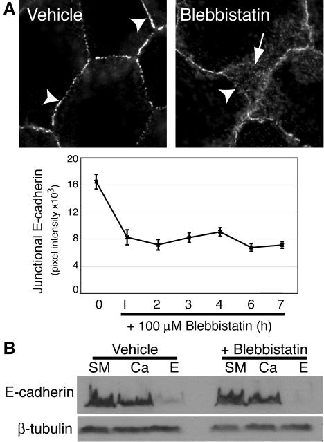
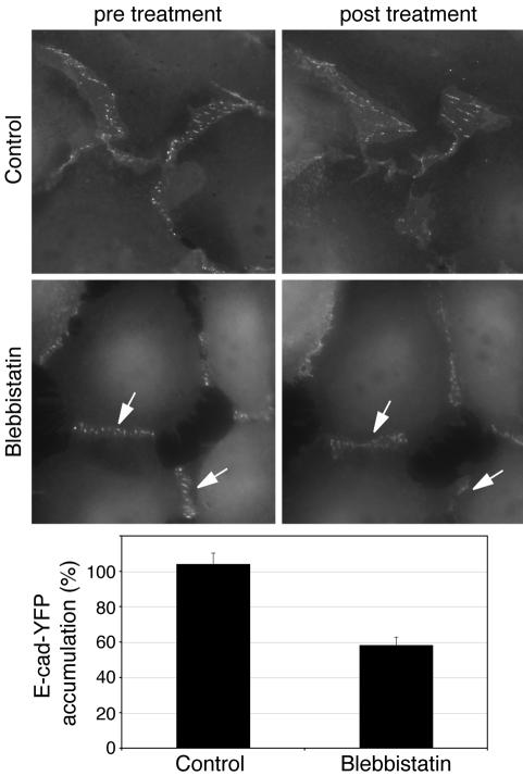
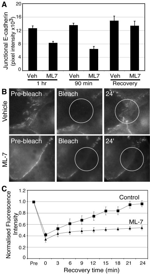
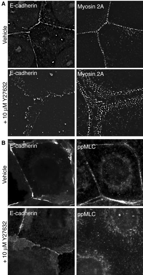
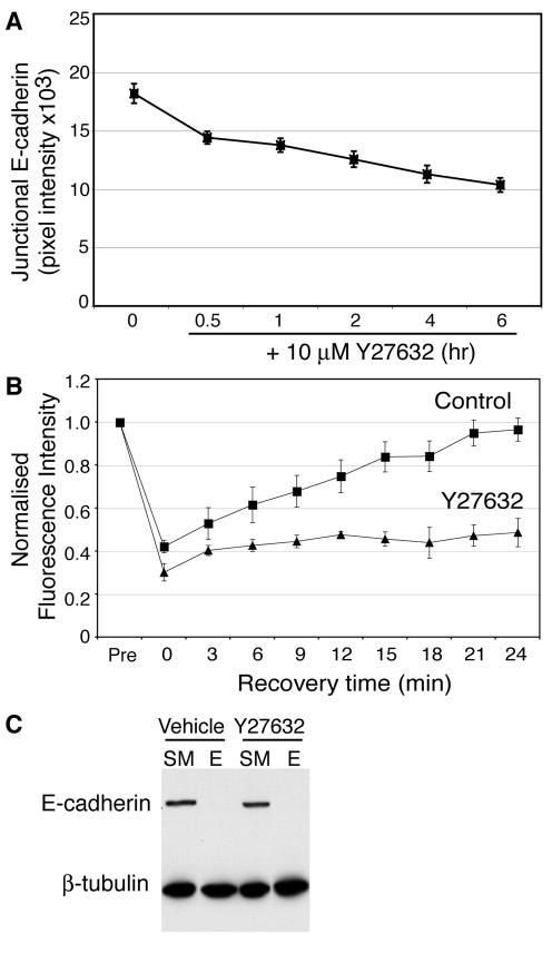
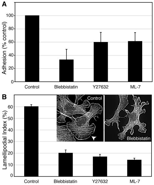
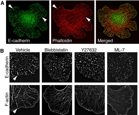
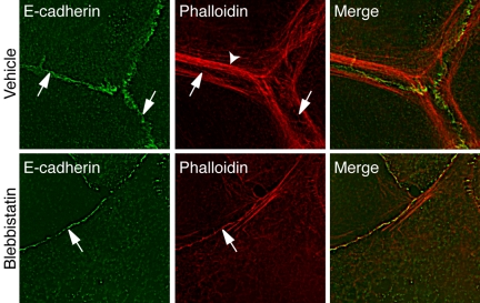
Classical cadherins accumulate at cell-cell contacts as a characteristic response to productive adhesive ligation. Such local accumulation of cadherins is a developmentally regulated process that supports cell adhesiveness and cell-cell cohesion. Yet the molecular effectors responsible for cadherin accumulation remain incompletely understood. We now report that Myosin 2 is critical for cells to concentrate E-cadherin at cell-cell contacts. Myosin 2 is found at cadherin-based cell-cell contacts and its recruitment requires E-cadherin activity. Indeed, both Myosin 2 recruitment and its activation were stimulated by E-cadherin homophilic ligation alone. Inhibition of Myosin 2 activity by blebbistatin or ML-7 rapidly impaired the ability of cells to concentrate E-cadherin at adhesive contacts, accompanied by decreased cadherin-based cell adhesiveness. The total surface expression of cadherins was unaffected, suggesting that Myosin 2 principally regulates the regional distribution of cadherins at the cell surface. The recruitment of Myosin 2 to cadherin contacts, and its activation, required Rho kinase; furthermore, inhibition of Rho kinase signaling effectively phenocopied the effects of Myosin 2 inhibition. We propose that Myosin 2 is a key effector of Rho-Rho kinase signaling that regulates cell-cell adhesion by determining the ability of cells to concentrate cadherins at contacts in response to homophilic ligation.


Figures

Figure 1.
Figure 1.
Myosin 2 accumulates at cell–cell contacts in an E-cadherin-dependent manner. (A) Endogenous Myosin 2A localizes in puncta at cadherin-based cell–cell contacts. Confluent MCF-7 monolayers were fixed and labeled with antibodies specific for E-cadherin and Myosin 2A. (B) Exogenous GFP-tagged Myosin 2A also localizes in regions within E-cadherin-based cell–cell contacts. MCF-7 cells were transiently transfected with EGFP-tagged Myosin 2A, briefly extracted with TX-100 before fixation and costained both for E-cadherin and the GFP epitope tag (GFP-Myosin 2A). A single image plane is shown after 3D deconvolution. (C) E-cadherin is necessary for Myosin 2A localization at cell–cell contacts. Confluent MCF-7 cell monolayers were incubated in medium alone (No antibody) or in the presence of the E-cadherin function-blocking SHE78–7 mAb (1:50) for 15 min. E-cadherin was detected using the prebound blocking antibody, whereas Myosin 2A was detected using isoform-specific antibodies. Exposure to SHE78–7 substantially reduced and reorganized Myosin 2A staining at contacts that still retained E-cadherin.
Figure 2.
Figure 2.
E-cadherin homophilic ligation is sufficient to recruit and activate Myosin 2A. (A) E-cadherin homophilic ligation triggers the recruitment of Myosin 2A to sites of adhesion. Latex beads coated with either hE/Fc or ConA were allowed to adhere for 90 min to the dorsal surface of hE-CHO cells transiently expressing GFP-tagged Myosin 2A (GFP-Myosin 2A). Cells were then immunolabeled with antibodies specific for GFP and β-catenin (marking the cadherincatenin complex). Both GFP-Myosin 2A and the cadherin complex show significantly greater accumulation in flares around the hE/Fc-beads (arrowheads) when compared with ConA-coated control beads. Incubation with either Y-27632 (10 μM) or ML-7 (10 μM) markedly reduced the recruitment of GFP-Myosin 2A to cadherin homophilic adhesions. (B) E-cadherin homophilic ligation activates Myosin 2. Cells were allowed to adhere to dishes coated with either poly-l-lysine (PLL) or hE/Fc in the presence of blebbistatin (Blebbi, 10 μM), Y-27632 (10 μM), or ML-7 (10 μM) for 90 min. Western blots from cell lysates were probed for di-phosphorylated, activated myosin light chain (ppMLC) or β-tubulin (as a loading control).
Figure 3.
Figure 3.
Myosin 2 motor activity is necessary for the local concentration of E-cadherin at cell–cell contacts. (A) Blebbistatin perturbs E-cadherin accumulation in cell–cell contacts. MCF-7 cells were treated with blebbistatin in complete growth media for varying periods of time and then fixed and processed for E-cadherin immunofluorescence. Representative images of cells treated with drug or control for 60 min are shown. Note that although vehicle-treated cells showed continuous chicken-wire E-cadherin staining (arrowheads), in drug-treated cells the apical cadherin staining was less intense and often discontinuous (arrowhead), whereas E-cadherin staining also extended away in overlapping cell–cell contacts. E-cadherin fluorescence intensity at cell–cell contacts (Junctional E-cadherin) was measured by digital image analysis of drug-treated and control cultures (data are means ± SE). (B) Inhibition of Myosin 2 does not affect the surface expression of E-cadherin. MCF-7 cells were treated with blebbistatin (100 μM) for 90 min before exposure to crystalline trypsin for 20 min in the presence of either 2 mM CaCl2 (Ca) or 5 mM EGTA (E). Lysates from these cells and untreated cells (SM, starting material) were subjected to SDS-PAGE and immunoblotted for E-cadherin and β-tubulin (as a loading control).
Figure 4.
Figure 4.
Blebbistatin perturbs the accumulation of YFP-tagged E-cadherin at cell–cell contacts. CHO cells stably expressing E-Cad-YFP were examined by live-cell imaging. Individual contacts were imaged (600-ms exposures) before, and 1 h after, addition of blebbistatin (10 μM) using identical camera acquisition settings. Fluorescence intensity at contacts was determined as described in Materials and Methods; fluorescence intensity after blebbistatin (E-Cad-YFP accumulation) was expressed as a percentage of fluorescence intensity before the drug; n = 10–14, p < 0.001 (Student's t test). E-Cad-YFP accumulated in prominent puncta at cell–cell contacts. Puncta were less prominent (arrows), and fluorescence intensity at contacts reduced, after treatment with blebbistatin.
Figure 5.
Figure 5.
Myosin light-chain kinase activity is necessary for local accumulation of E-cadherin at cell–cell contacts. (A) Inhibition of MLCK affects E-cadherin accumulation at cell–cell contacts. MCF-7 monolayers were incubated with ML-7 (10 μM) or vehicle for 60–90 min then processed for E-cadherin immunofluorescence and quantitation. One set of cells (Recovery) were treated with drug for 60 min and then allowed to recover in fresh drug-free medium before fixation. (B) ML-7 affects recovery of E-cadherin-YFP fluorescence after photobleaching of cell–cell contacts. Contacts between CHO cells stably expressing E-cadherin-YFP were photobleached, and the subsequent recovery of cadherin fluorescence was monitored by time-lapse epi-fluorescence microscopy. Cells were incubated with ML-7 (10 μM) or vehicle alone before experiments. Representative images from movies are shown; circles mark the photobleached areas. (C) E-cadherin-YFP fluorescence recovery at cell–cell contacts after photobleaching was calculated and modeled as detailed in Materials and Methods.
Figure 6.
Figure 6.
Myosin 2 and activated MLC localizes to cell–cell contacts in response to Rho kinase signaling. MCF-7 monolayers were treated with 10 μM Y-27632 for 30 min, fixed, and processed for immunolocalization of endogenous Myosin 2A and E-cadherin (A) or ppMLC and E-cadherin (B). Strikingly, Y-27632 treatment reduced significantly the accumulation of both Myosin 2 and ppMLC at cell–cell contacts.
Figure 7.
Figure 7.
Rho kinase signaling is necessary for the local accumulation of E-cadherin at cell–cell contacts. (A) Accumulation of endogenous E-cadherin at contacts between MCF-7 cells treated with Y-27632 (10 μM) for 0–6 h. E-cadherin accumulation at cell–cell contacts was measured by quantitative immunofluorescence microscopy. Data are means ± SE. (B) FRAP analysis of E-cadherin-YFP accumulation at contacts between hE-YFP-CHO cells treated with either Y-27632 (10 μM) or vehicle alone. (C) Surface expression of E-cadherin in MCF-7 cells treated with Y-27632 (10 μM) or vehicle alone was measured by surface trypsin protection assays (SM, starting material, E, trypsinization in the presence of EGTA). All the cadherin remained accessible to surface trypsinization in both control and drug-treated cells. These data were representative of three independent experiments.
Figure 8.
Figure 8.
Myosin 2 activity is necessary for cadherin homophilic adhesion. (A) Inhibition of Myosin 2 activity affects E-cadherin-mediated adhesion measured by resistance to detachment from cadherin-coated substrata. hE-CHO cells were plated onto substrata coated with hE/Fc and allowed to adhere in the presence or absence of blebbistatin (10 μM), Y-27632 (10 μM), or ML-7 (10 μM) for 90 min. Cells were then detached and residual adherent cells measured as described in Materials and Methods. The numbers of adherent cells in drug-treated samples were normalized to controls subjected to the same experimental manipulations. Experiments were performed in triplicate on at least three separate occasions. (B) Myosin activity is necessary for E-cadherin-mediated contact zone extension. Freshly isolated hE-CHO cells were allowed to adhere to and extend contacts upon hE/Fc-coated substrata for 90 min in the presence of blebbistatin (10 μM), Y-27632 (10 μM), or ML-7 (10 μM). The ability of cells to extend contacts (Lamellipodial Index) was measured on phalloidin-stained samples as described in Materials and Methods. Data are means ± SE (n = 30–35). Insets show representative images of phalloidin-stained control and blebbistatin-treated cells; the arrowhead indicates a broad cadherin-based lamella.
Figure 9.
Figure 9.
Myosin 2 activity supports cadherin-based actin bundles in homophilic adhesion assays. (A) Two patterns of F-actin organization are evident in hE-CHO cells adherent to hE/Fc-coated substrata. Cells were allowed to adhere to hE/Fc for 90 min before fixation and immunostaining for cellular E-cadherin using antibodies directed against the cytoplasmic tail and F-actin (phalloidin). Phalloidin staining revealed dense bands of F-actin at the outer margins of cadherin-dependent lamellipodia and also prominent bundles that typically terminated in large cadherin clusters (macroclusters, arrowheads). (B) Lateral organization of cellular E-cadherin and formation of actin bundles at cadherin adhesive interfaces requires Myosin 2. hE-CHO cells that had adhered to hE/Fc-coated coverslips for 60 min were then treated with the indicated drugs (all at 10 μM) for a further 30 min. Cadherin macroclusters (arrowheads) and the actin bundles they demarcate were largely abolished in drug-treated cells.
Figure 10.
Figure 10.
Blebbistatin perturbs perijunctional actin cables. Confluent MCF-7 monolayers were stained for E-cadherin or F-actin as indicated. In control cells F-actin was found in prominent perijunctional actin cables (arrowheads) as well as in fine cortical bands (arrows) that localized with E-cadherin itself (arrows). Blebbistatin (10 μM, 90 min) abolished many of the perijunctional actin bundles, but residual cortical F-actin staining was evident at the cell–cell contacts (arrows).

References

    1. Adams, C. L., Chen, Y.-T., Smith, S. J., and Nelson, W. J. (1998). Mechanisms of epithelial cell–cell adhesion and cell compaction revealed by high-resolution tracking of E-cadherin-green fluorescent protein. J. Cell Biol. 142, 1105–1119. - PMC - PubMed
    1. Amano, M., Ito, M., Kimura, K., Fukata, Y., Chihara, K., Nakano, T., Matsuura, Y., and Kaibuchi, K. (1996). Phosphorylation and activation of myosin by Rho-associated kinase (Rho-kinase). J. Biol. Chem. 271, 20246–20249. - PubMed
    1. Avizienyte, E., Fincham, V. J., Brunton, V. G., and Frame, M. C. (2004). Src SH3/2 domain-mediated peripheral accumulation of Src and phospho-myosin is linked to deregulation of E-cadherin and the epithelial-mesenchymal transition. Mol. Biol. Cell 15, 2794–2803. - PMC - PubMed
    1. Bertet, C., Sulak, L., and Lecuit, T. (2004). Myosin-dependent junction remodelling controls planar cell intercalation and axis elongation. Nature 429, 667–671. - PubMed
    1. Boggon, T. J., Murray, J., Chappuis-Flament, S., Wong, E., Gumbiner, B. M., and Shapiro, L. (2002). C-cadherin ectodomain structure and implications for cell adhesion mechanisms. Science 296, 1308–1313. - PubMed

Publication types

LinkOut - more resources