. 2005 Oct;16(10):4543-56.
doi: 10.1091/mbc.e05-03-0243. Epub 2005 Jul 19.

Molecular interactions position Mso1p, a novel PTB domain homologue, in the interface of the exocyst complex and the exocytic SNARE machinery in yeast

Affiliations

Molecular interactions position Mso1p, a novel PTB domain homologue, in the interface of the exocyst complex and the exocytic SNARE machinery in yeast

Michael Knop et al. Mol Biol Cell. 2005 Oct.

Abstract

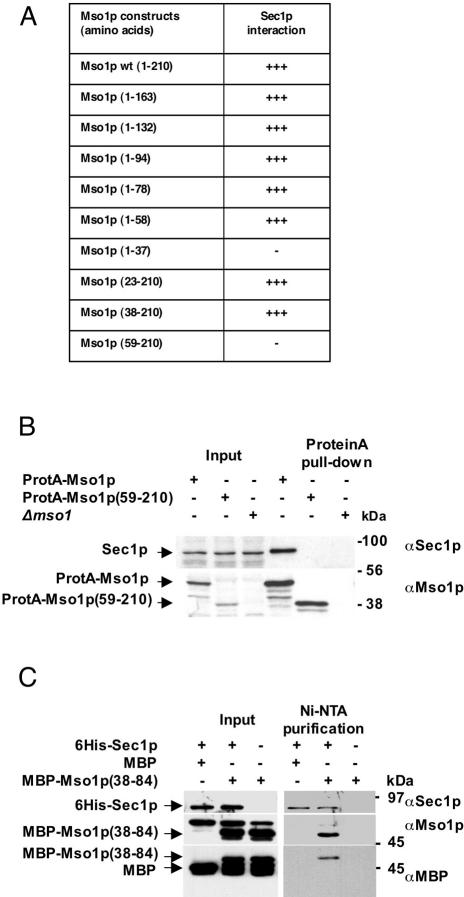
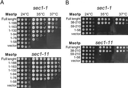
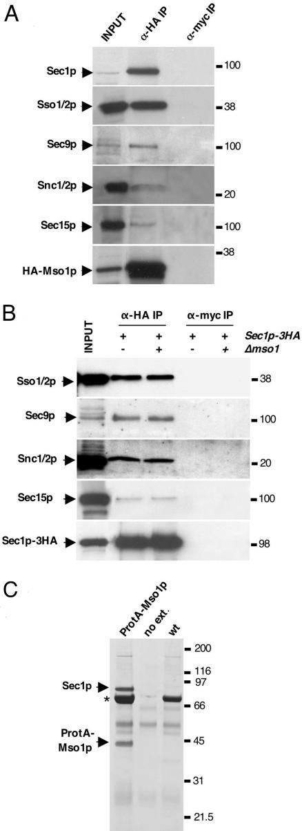
In this study, we have analyzed the association of the Sec1p interacting protein Mso1p with the membrane fusion machinery in yeast. We show that Mso1p is essential for vesicle fusion during prospore membrane formation. Green fluorescent protein-tagged Mso1p localizes to the sites of exocytosis and at the site of prospore membrane formation. In vivo and in vitro experiments identified a short amino-terminal sequence in Mso1p that mediates its interaction with Sec1p and is needed for vesicle fusion. A point mutation, T47A, within the Sec1p-binding domain abolishes Mso1p functionality in vivo, and mso1T47A mutant cells display specific genetic interactions with sec1 mutants. Mso1p coimmunoprecipitates with Sec1p, Sso1/2p, Snc1/2p, Sec9p, and the exocyst complex subunit Sec15p. In sec4-8 and SEC4I133 mutant cells, association of Mso1p with Sso1/2p, Snc1/2p, and Sec9p is affected, whereas interaction with Sec1p persists. Furthermore, in SEC4I133 cells the dominant negative Sec4I133p coimmunoprecipitates with Mso1p-Sec1p complex. Finally, we identify Mso1p as a homologue of the PTB binding domain of the mammalian Sec1p binding Mint proteins. These results position Mso1p in the interface of the exocyst complex, Sec4p, and the SNARE machinery, and reveal a novel layer of molecular conservation in the exocytosis machinery.


Figures

Figure 1.
Figure 1.
Initiation of de novo plasma membrane biogenesis at the SPBs is blocked in Δmso1 mutant cells. (A) Wide field immunofluorescence pictures of representative cells of wt (YKS32), Δmpc54 Δmpc70 (YKS65-1), and Δmso1 (MBY13-1-1) strains in metaphase (top) and in anaphase of meiosis II (bottom). DAPI staining of DNA (blue), Ady3p (red), and tubulin (green) are shown. Some cytoplasmic and SPB-associated precursor structures (white open triangles in metaphase cells, yellow arrows in anaphase cells) and the leading edge protein coats (white arrows) are indicated. In anaphase of meiosis II (A, bottom), Ady3p accumulates at the spindle pole in the Δmso1 cells (yellow arrows), whereas in wild-type cells it localizes to the leading edge protein coat of the prospore membranes (outlined with yellow dotted lines). In Δmpc54 Δmpc70 cells, only marginal accumulation of Ady3p at the SPB is observed. Bar (A), 2 μm. (B) Electron microscopy analysis of PSM formation in wt, Δmcp54 Δmcp70, and Δmso1 mutants. White arrows point to the inner side of the SPBs, and asterisks mark the meiotic plaque of the SPB. White triangles outline assembled prospore membranes. Δmcp54 Δmcp70 cells are devoid of the meiotic plaque, and only a few vesicles are found in the vicinity of the SPB. A population of homogenous unfused vesicles (open triangles) is found attached to the meiotic SPBs in Δmso1 mutants.
Figure 2.
Figure 2.
Localization of Mso1–4GFP in mitotic and meiotic cells. Vegetatively growing living haploid cells (YJM2) (A) and fixed sporulating diploid cells (YMK799) in meiosis (B) expressing chromosomally encoded MSO1–4GFP at endogenous expression levels were investigated by fluorescence microscopy. (A) Mso1–4GFP localized to the area of bud formation in an unbudded cell (arrow) and at the tip of the bud (open triangle) and the bud neck (arrow heads) in budded cells. (B) Immunofluorescence localization of Mso1–4GFP to small prospore membranes in fixed meiotic cells in metaphase (top) and in anaphase (bottom). The size of the prospore membranes is indicated by codetection of Ady3p at the leading edge of the prospore membrane, whereas tubulin staining depicts the positions of the meiotic spindles. Bar, 1 μm (A) and 2 μm (B).
Figure 3.
Figure 3.
Mapping of the Mso1p domain responsible for Sec1p interaction. (A) Table summarizing yeast two-hybrid interactions of Mso1p mutants with Sec1p. The “+” indicate the relative strength of the interaction based on β-galactosidase activity and growth on medium lacking leucine. (B) The amino terminus of Mso1p is needed for Sec1p interaction in yeast cell lysates. Protein A pull-downs with IgG-coupled beads were performed with cell lysates expressing ProtA-Mso1p (H2937), ProtA-Mso1p(59-210) (H2955), or lysates deleted for Mso1p (Δmso1) (H2785). The input and pull-down samples were analyzed by Western blotting using anti-Sec1p and Mso1p antibodies. (C) In vitro binding of His6-Sec1p with its binding domain in Mso1p(38-84). Only in samples containing His6-Sec1p and MBP-Mso1p(38-84) copurification is detected. In negative control samples, no His6-Sec1p and MBP copurification is observed. Likewise, no copurification of MBP-Mso1p(38-84) with Ni-NTA resin alone is observed. Positions of molecular weight standards are indicated on the right. Antibodies to Mso1p, MBP, or Sec1p were used for protein detection.
Figure 4.
Figure 4.
The Sec1p binding domain is necessary, but not sufficient for Mso1p in vivo function. Temperature-sensitive sec1-1 (H305) and sec1-11 (H306) mutants were transformed with high-copy plasmids harboring wt MSO1 or fragments of MSO1. The growth of the transformants was scored at different temperatures. (A) The effect of carboxy-terminal truncations of Mso1p. (B) The effect of amino-terminal truncations of Mso1p. The amino acids encoded by the fragments are indicated by numbers in the figures. The growth of serial 10-fold dilutions of transformed cells was monitored at the temperatures indicated in the figures.
Figure 5.
Figure 5.
The mso1T47A mutation abolishes Mso1p in vivo function and displays specific genetic interactions. (A) The point mutation T47A within the Sec1p binding domain abolishes the sec1 mutant suppression activity of Mso1p. (B) Table summarizing synthetic lethal interactions between different mso1-mutants and sec1, sec2, and sec4 mutants. A “+” indicates a synthetic lethal interaction, and “–” indicates a viable combination of the mutations.
Figure 6.
Figure 6.
Immunoprecipitations analysis of Mso1p associating protein complexes. (A) Mso1p coimmunoprecipitates with Sec1p, the Snc1/2p–Sec9p–Sso1/2p SNARE complex subunits and Sec15p. Anti-HA immunoprecipitations were carried out from yeast cell (H2657) lysates where the sole copy of cellular Mso1p is triple HA-tagged at the carboxy terminus. Analysis of the immunoprecipitates was carried out by Western blotting with antibodies to HA, Sec1p, Sso1/2p, Sec9p, Snc1/2p, and Sec15p. (B) Effect of Mso1p deletion on the Sec1p, Snc1/2p, Sec9p, Sso1/2p, and Sec15p coimmunoprecipitation. Anti-HA immunoprecipitations were carried out in yeast strains expressing Sec1p-3HA in the presence (YNR53-1) or absence (YNR60-3) of Mso1p. The precipitates were analyzed for the presence of SNARE molecules and Sec15p with specific antibodies indicated on the left. Deletion of Mso1p does not affect the composition of the complex. (C) Sec1p copurifies in equimolar manner with ProtA-Mso1p (Coomassie staining). Protein A complex purification from cells expressing Protein A-Mso1p (YAM274-2). wt, cells expressing untagged Mso1p (YKS32); no ext., only IgG-beads without cell extract. Positions of molecular weight standards are indicated on the right. Asterisk marks the position of the copurifying, nonspecific virus L-A coat protein.
Figure 7.
Figure 7.
Immunoblot analysis of Mso1p-3HA immunoprecipitates in different sec4 mutant expressing cells. (A) In the experiment, strains expressing either Sec2p-3HA (YNR27) or Mso1p-3HA (H2657) (both from their endogenous promoter) or no HA-tagged protein (EMS356-1) (as indicated above the blots), and their variants, where chromosomally integrated dominant alleles of SEC4 (S34N or N133I) under the control of the GAL1 promoter, were used. On induction of the GAL1 promoter for 2 h, cell extracts were subjected to anti-HA immunoprecipitations. Isolated immunocomplexes were separated on 4–12% Bis-Tris NU PAGE gradient gels followed by immunoblotting with the indicated antibodies (anti-HA IP). Extracts were analyzed for the presence of the expressed sec4-alleles (extract). More than 90% of HA-tagged proteins present in the crude extracts were precipitated (our unpublished data). (B) The effect of sec4-8 mutation on Mso1p protein associations. sec4-8 cells (H3331) expressing Mso1p-3HA from MSO1 promoter were incubated either at 24 or 37°C, lysed and subjected for anti-HA immunoprecipitations. The presence of indicated proteins was analyzed by Western blotting. Coimmunoprecipitation of Mso1p-3HA with Sso and Sec9 proteins is affected already at 24°C, whereas Mso1p association with Sec1p and Sec15p is not affected.
Figure 8.
Figure 8.
Mso1p is homologous to PTB domains from Mint1 and Mint2. (A) Comparison of S. cerevisiae (Sc) Mso1p with homologues found in Saccharomyces mikatae (Sm), K. lactis (Kl), and Candida albicans (Ca). (B) Comparison of KlMso1p with PTB domains of human HsMint1 and HsMint2. (C) Phylogenetic tree of the PTB domains of Homo sapiens (Hs) Mint 1, 2, and 3, and Mso1p homologues in S. pombe (Sp), K. lactis (Kl), C. albicans (Ca), Saccharomyces castellii (Sca), S. cerevisiae (Sc), and S. mikatae (Sm). The scale equals the distance where 0.1 nucleotide substitutions per site have occurred. In the pileups, the asterisk (*) marks identical, colon (:), strongly similar, and period (.), similar amino acids.

Similar articles

Cited by

References

    1. Aalto, M. K., Jantti, J., Ostling, J., Keranen, S., and Ronne, H. (1997). Mso1p: a yeast protein that functions in secretion and interacts physically and genetically with Sec1p. Proc. Natl. Acad. Sci. USA 94, 7331–7336. - PMC - PubMed
    1. Banta, L. M., Vida, T. A., Herman, P. K., and Emr, S. D. (1990). Characterization of yeast Vps33p, a protein required for vacuolar protein sorting and vacuole biogenesis. Mol. Cell. Biol. 10, 4638–4649. - PMC - PubMed
    1. Biederer, T., and Sudhof, T. C. (2000). Mints as adaptors. Direct binding to neurexins and recruitment of munc18. J. Biol. Chem. 275, 39803–39806. - PubMed
    1. Borg, J. P., Ooi, J., Levy, E., and Margolis, B. (1996). The phosphotyrosine interaction domains of X11 and FE65 bind to distinct sites on the YENPTY motif of amyloid precursor protein. Mol. Cell. Biol. 16, 6229–6241. - PMC - PubMed
    1. Borg, J. P., Straight, S. W., Kaech, S. M., de Taddeo-Borg, M., Kroon, D. E., Karnak, D., Turner, R. S., Kim, S. K., and Margolis, B. (1998). Identification of an evolutionarily conserved heterotrimeric protein complex involved in protein targeting. J. Biol. Chem. 273, 31633–31636. - PubMed

Publication types

MeSH terms