. 2005 Nov 15;392(Pt 1):173-80.
doi: 10.1042/BJ20050354.

Haem repression of the housekeeping 5-aminolaevulinic acid synthase gene in the hepatoma cell line LMH

Affiliations

Haem repression of the housekeeping 5-aminolaevulinic acid synthase gene in the hepatoma cell line LMH

Sridevi Kolluri et al. Biochem J. .

Abstract

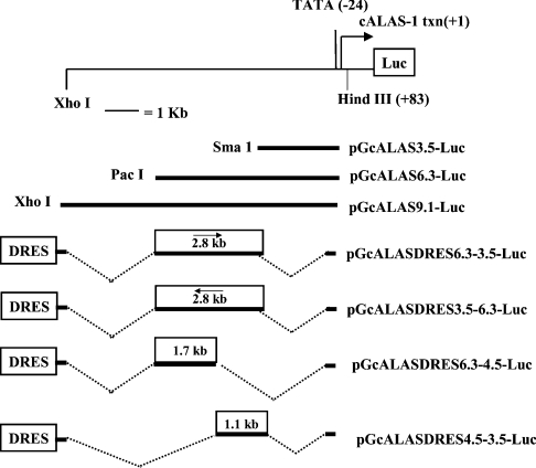
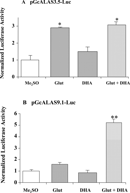
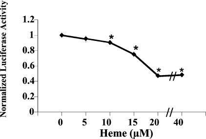
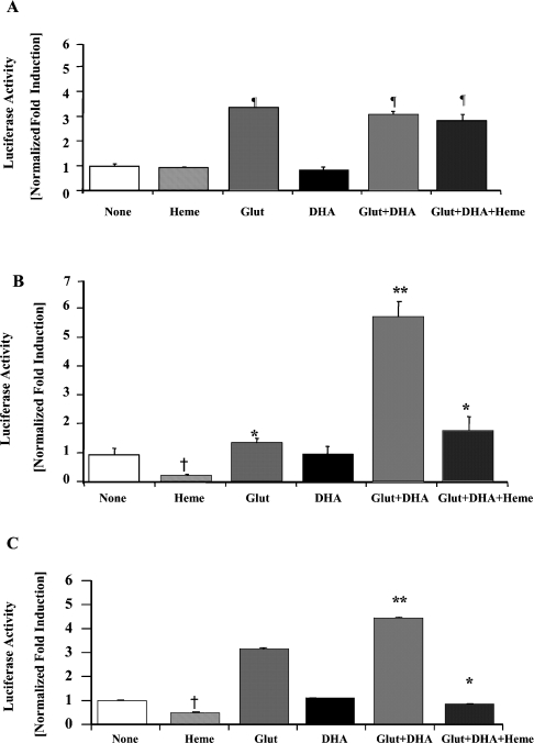
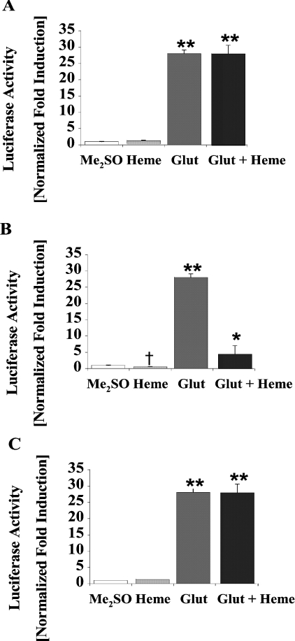
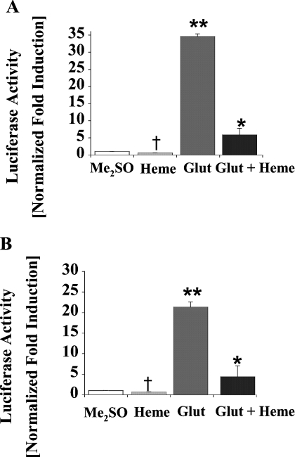
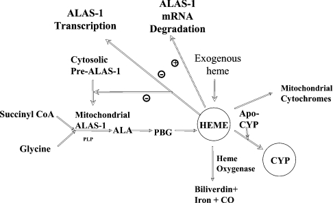
Haem is essential for the health and function of nearly all cells. 5-Aminolaevulinic acid synthase-1 (ALAS-1) catalyses the first and rate-controlling step of haem biosynthesis. ALAS-1 is repressed by haem and is induced strongly by lipophilic drugs that also induce CYP (cytochrome P450) proteins. We investigated the effects on the avian ALAS-1 gene promoter of a phenobarbital-like chemical, Glut (glutethimide), and a haem synthesis inhibitor, DHA (4,6-dioxoheptanoic acid), using a reporter gene assay in transiently transfected LMH (Leghorn male hepatoma) hepatoma cells. A 9.1 kb cALAS-1 (chicken ALAS-1) promoter-luciferase-reporter construct, was poorly induced by Glut and not by DHA alone, but was synergistically induced by the combination. In contrast, a 3.5 kb promoter ALAS-1 construct was induced by Glut alone, without any further effect of DHA. In addition, exogenous haem (20 microM) repressed the basal and Glut- and DHA-induced activity of luciferase reporter constructs containing 9.1 and 6.3 kb of ALAS-1 5'-flanking region but not the construct containing the first 3.5 kb of promoter sequence. This effect of haem was subsequently shown to be dependent on the -6.3 to -3.5 kb region of the 5'-flanking region of cALAS-1 and requires the native orientation of the region. Two deletion constructs of this approx. 2.8 kb haem-repressive region (1.7 and 1.1 kb constructs) retained haem-dependent repression of basal and drug inductions, suggesting that more than one cis-acting elements are responsible for this haem-dependent repression of ALAS-1. These results demonstrate that there are regulatory regions in the 5'-flanking region of the cALAS-1 gene that respond to haem and provide a basis for further investigations of the molecular mechanisms by which haem down-regulates expression of the ALAS-1 gene.


Figures

Figure 1
Figure 1. Partial restriction map of the 5′-UTR and 5′-flanking region of the cALAS-1 gene, and structures of cALAS-1 constructs
Numbering starts at the transcription start point (+1). Recognition sites for restriction endonucleases XhoI, PacI, SmaI and HindIII used for plasmid construction are shown. The portions of the 5′-flanking region and 5′-UTR cloned into the reporter (Luc) gene plasmids (pGL3-Basic or pGL3-Promoter) are indicated, as are the abbreviations used to denote these constructs. A shorter construct, composed of the first 299 bp upstream of the transcription starting point, linked to the luciferase gene, was also studied. It showed no effect of up- or down-regulation.
Figure 2
Figure 2. Effects of Glut and DHA on pGcALAS3.5-Luc (A) or pGcALAS9.1-Luc (B) transiently transfected into LMH cells
Following transfection, LMH cells were treated with vehicle (Me2SO), Glut (50 μM), or DHA (250 μM), alone or in combination, for 16 h prior to harvest. Luciferase and β-gal activities and protein concentrations were assayed as described in the Materials and methods section. Luciferase activities are expressed as fold induction compared with vehicle-treated control, normalized to β-gal activity and protein concentrations. Results are means±S.E.M. for triplicate determinations, and representative of six independent experiments. *Differs from Me2SO or DHA alone, P<0.01. In (A), the two groups treated with Glut did not differ significantly from one another. **Differs from all others, P<0.001.
Figure 3
Figure 3. Effect of exogenous haem on pGcALAS9.1-Luc activity
LMH cells were transiently transfected with pGcALAS9.1-Luc and treated with increasing concentrations of haem (‘Heme’; 0, 5, 10, 15, 20 and 40 μM) dissolved in Me2SO. After 16 h, the cells were harvested and lysates were used to measure luciferase, β-gal and protein levels, as described in the Materials and methods section. Results are means±S.E.M. for triplicate determinations, and are representative of four independent experiments. Luciferase activities are expressed as fold induction compared with vehicle-treated control, normalized to β-gal and protein levels. The amount of Me2SO added per well was kept constant and was not greater than 2 μl/ml medium, a volume that was shown in previous studies to not affect luciferase activity [26,28,45,46]. *Differs from cells transfected with pGcALAS9.1-Luc and treated with 0 μM haem, P<0.001.
Figure 4
Figure 4. Effects of Glut, DHA and haem in LMH cells transiently transfected with pGcALAS3.5-Luc (A), pGcALAS9.1-Luc (B) or pGcALAS6.3-Luc (C)
Transfected cells were harvested 16 h after chemical treatment, and lysates were used to measure luciferase, β-gal and protein levels. Concentrations of the chemicals were as follows: Glut, 50 μM; DHA, 250 μM; and haem, 20 μM. Results are means±S.E.M. for triplicate determinations, and representative of five independent experiments. Luciferase activities are expressed as fold induction compared with vehicle-treated control, normalized to β-gal and protein levels. *Differs from Glut+DHA, P<0.001. **Differs from others, P<0.001. ¶Differs from Me2SO, haem or DHA alone, P<0.01. In (A), the three groups treated with Glut did not differ significantly from one another. †Significant decrease from cells treated with Me2SO, P<0.001.
Figure 5
Figure 5. Effects of Glut and haem on LMH cells transiently transfected with pGL3DRESLuc (A), pGcALASDRES6.3-3.5-Luc (B) or pGcALASDRES3.5-6.3-Luc (C)
Transfected cells were treated, harvested and assayed as described in the Materials and methods section and the legend to Figure 4. Results are means±S.E.M. for triplicate determinations and representative of five independent experiments. **Glut differs significantly from vehicle (Me2SO) alone or haem alone, P<0.001; *Glut + Haem differs significantly from Glut alone, P<0.001. †Significant decrease from cells treated with Me2SO, P<0.01.
Figure 6
Figure 6. Effects of Glut, DHA and haem on LMH cells transiently transfected with pGcDRES 6.3-4.5-Luc (A) or pGL3 DRES 4.5-3.5-Luc (B)
Transfected cells were treated, harvested and assayed as described in the Materials and methods section and the legend to Figure 4. Results are means±S.E.M. for triplicate determinations and representative of five independent experiments. †Differs from vehicle alone (Me2SO) or Glut + Haem, P<0.01. **Differs from all other treatment groups, P<0.0001. *Differs from Glut alone, P<0.001.
Figure 7
Figure 7. Effects of the regulatory haem pool on hepatic haem metabolism
Haem exerts regulatory effects on both ALAS-1 and HO-1 to control both its synthesis and degradation. ALAS-1 catalyses the formation of ALA from glycine and succinyl-CoA. Haem is synthesized from ALA through a series of intermediary steps catalysed by the enzymes of the haem biosynthetic pathway. Several steps in the production of functional mature ALAS-1 protein are down-regulated by exogenous or endogenous haem, as indicated by the minus signs, or by the plus sign indicating increased degradation of ALAS-1 mRNA. Haem is also incorporated into apo-haemoproteins [such as CYP (cytochrome P450) proteins and mitochondrial cytochromes] as the prosthetic group to form functional holo-haemoproteins.

Similar articles

Cited by

References

    1. Ades I. Z. Heme production in animal tissues: the regulation of biogenesis of delta-aminolevulinate synthase. Int. J. Biochem. 1990;22:565–578. - PubMed
    1. Bonkovsky H. L. Porphyrin and heme metabolism and the porphyrias. In: Zakim D., Boyer T., editors. Hepatology: A Textbook of Liver Disease. Philadelphia, PA: W.B. Saunders; 1990. pp. 378–424.
    1. May B. K., Dogra S. C., Sadlon T. J., Bhasker C. R., Cox T. C., Bottomley S. S. Molecular regulation of heme biosynthesis in higher vertebrates. Prog. Nucleic Acid Res. Mol. Biol. 1995;51:1–51. - PubMed
    1. Kappas A., Sassa S., Galbraith R. A., Nordmann Y. The porphyrias. In: Scriver C. R., Beaudet A. L., Sly W. S., Valle D., editors. The Metabolic Basis of Inherited Disease. New York: McGraw-Hill; 1995. pp. 2103–2160.
    1. Riddle R. D., Yamamoto M., Engel J. D. Expression of delta-aminolevulinate synthase in avian cells: separate genes encode erythroid-specific and nonspecific isozymes. Proc. Natl. Acad. Sci. U.S.A. 1989;86:792–796. - PMC - PubMed

Publication types

MeSH terms