

Overcoming cisplatin resistance by mTOR inhibitor in lung cancer
- PMID: 16033649
- PMCID: PMC1181826
- DOI: 10.1186/1476-4598-4-25
Overcoming cisplatin resistance by mTOR inhibitor in lung cancer
Abstract
Background: Cisplatin resistance is complex and involves several different mechanisms. Employing cDNA microarray analysis, we have found that cisplatin resistant cells share the common characteristic of increase in ribosomal proteins and elongation factors. We hypothesize that in order to survive cisplatin treatment, cells have to synthesize DNA repair proteins, antiapoptotic proteins and growth-stimulating proteins. Thus, by blocking the translation of these proteins, one should be able to restore cisplatin sensitivity. We have studied the role of CCI-779, an ester analog of rapamycin which is known to inhibit translation by disabling mTOR, in restoring cisplatin sensitivity in a panel of cisplatin resistant cell lines. We have also determined the role of CCI-779 in P-gp1 and MRP1 mediated resistance.
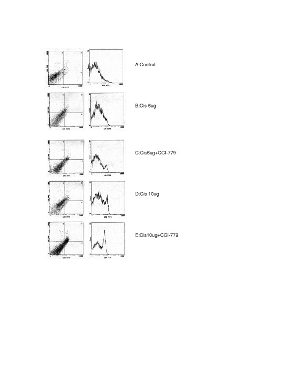
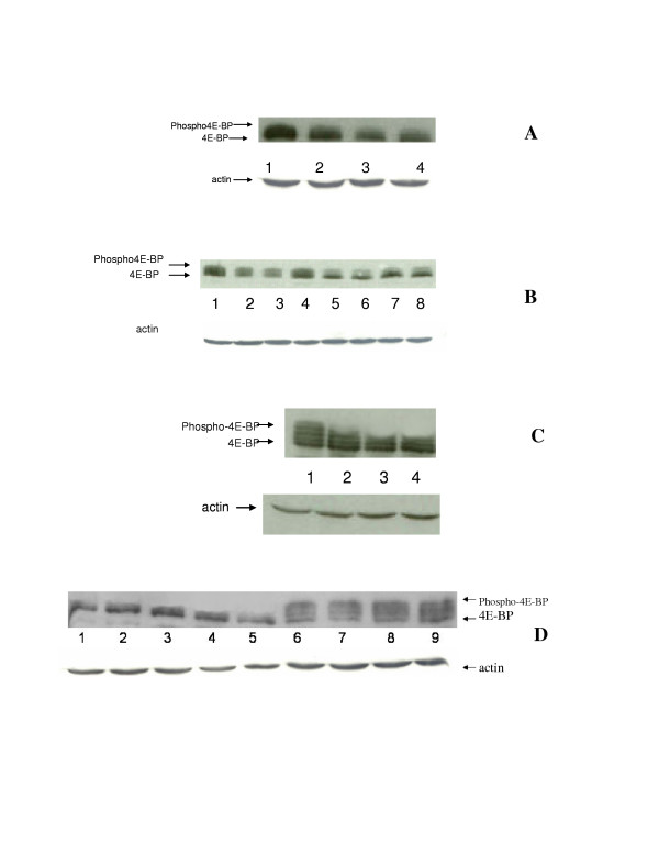
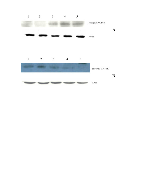
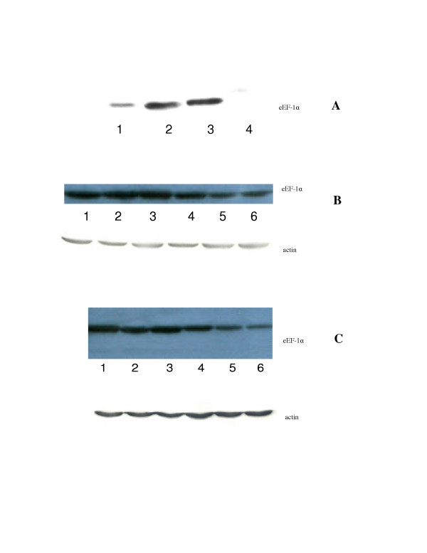
Results: Our data show that CCI-779 possess antiproliferative effects in both cisplatin sensitive and resistant cell lines, but shows no effect in P-gp1 and MRP1 overexpressing cell lines. Importantly, CCI-779 at 10 ng/ml (less that 10% of the growth inhibitory effect) can increase the growth inhibition of cisplatin by 2.5-6 fold. Moreover, CCI-779 also enhances the apoptotic effect of cisplatin in cisplatin resistant cell lines. In these resistant cells, adding CCI-779 decreases the amount of 4E-BP phosphorylation and p-70S6 kinase phosphorylation as well as lower the amount of elongation factor while cisplatin alone has no effect. However, CCI-779 can only reverse P-gp mediated drug resistance at a higher dose(1 ug/ml).
Conclusion: We conclude that CCI-779 is able to restore cisplatin sensitivity in small cell lung cancer cell lines selected for cisplatin resistance as well as cell lines derived from patients who failed cisplatin. These findings can be further explored for future clinical use. On the other hand, CCI-779 at achievable clinical concentration, has no growth inhibitory effect in P-gp1 or MRP1 overexpressing cells. Furthermore, CCI-779 also appears to be a weak MDR1 reversal agent. Thus, it is not a candidate to use in MDR1 or MRP1 overexpressing cells.
Figures

Similar articles
-
Rapamycin enhanced the antitumor efficacy of oxaliplatin in cisplatin-resistant ovarian cancer cells A2780cis both in vitro and in vivo.J Chemother. 2015;27(6):358-64. doi: 10.1179/1973947815Y.0000000021. Epub 2015 May 15. J Chemother. 2015. PMID: 25976336
-
CCI-779 (Temsirolimus) exhibits increased anti-tumor activity in low EGFR expressing HNSCC cell lines and is effective in cells with acquired resistance to cisplatin or cetuximab.J Transl Med. 2015 Apr 1;13:106. doi: 10.1186/s12967-015-0456-6. J Transl Med. 2015. PMID: 25890004 Free PMC article.
-
Enhanced sensitivity of multiple myeloma cells containing PTEN mutations to CCI-779.Cancer Res. 2002 Sep 1;62(17):5027-34. Cancer Res. 2002. PMID: 12208757
-
The rapamycin-sensitive signal transduction pathway as a target for cancer therapy.Oncogene. 2000 Dec 27;19(56):6680-6. doi: 10.1038/sj.onc.1204091. Oncogene. 2000. PMID: 11426655 Review.
-
Clinical development of mammalian target of rapamycin inhibitors.Hematol Oncol Clin North Am. 2002 Oct;16(5):1101-14. doi: 10.1016/s0889-8588(02)00051-5. Hematol Oncol Clin North Am. 2002. PMID: 12512385 Review.
Cited by
-
Enhanced Antitumor Activity with Combining Effect of mTOR Inhibition and Microtubule Stabilization in Hepatocellular Carcinoma.Int J Hepatol. 2013;2013:103830. doi: 10.1155/2013/103830. Epub 2013 Feb 20. Int J Hepatol. 2013. PMID: 23509629 Free PMC article.
-
Origins, genetic landscape, and emerging therapies of small cell lung cancer.Genes Dev. 2015 Jul 15;29(14):1447-62. doi: 10.1101/gad.263145.115. Genes Dev. 2015. PMID: 26220992 Free PMC article. Review.
-
Domain-based biosensor assay to screen for epidermal growth factor receptor modulators in live cells.Assay Drug Dev Technol. 2012 Feb;10(1):24-36. doi: 10.1089/adt.2011.423. Epub 2012 Jan 26. Assay Drug Dev Technol. 2012. PMID: 22280060 Free PMC article.
-
Inhibitors of mTOR.Oncologist. 2010;15(12):1262-9. doi: 10.1634/theoncologist.2010-0196. Epub 2010 Dec 8. Oncologist. 2010. PMID: 21147869 Free PMC article. Review.
-
Targeting of protein translation as a new treatment paradigm for prostate cancer.Curr Opin Oncol. 2017 May;29(3):210-220. doi: 10.1097/CCO.0000000000000367. Curr Opin Oncol. 2017. PMID: 28282343 Free PMC article.
References
-
- Vincent T. DeVita SHSAR. In: Cancer: Principles and Practice of Oncology Single Volume. 6th , editor. Vol. 1 2001.
-
- Lee KB, Parker RJ, Bohr V, Cornelison T, Reed E. Cisplatin sensitivity/resistance in UV repair-deficient Chinese hamster ovary cells of complementation groups 1 and 3. Carcinogenesis. 1993;14:2177–2180. - PubMed
-
- Murata T, Haisa M, Uetsuka H, Nobuhisa T, Ookawa T, Tabuchi Y, Shirakawa Y, Yamatsuji T, Matsuoka J, Nishiyama M, Tanaka N, Naomoto Y. Molecular mechanism of chemoresistance to cisplatin in ovarian cancer cell lines. Int J Mol Med. 2004;13:865–868. - PubMed
-
- Asselin E, Mills GB, Tsang BK. XIAP regulates Akt activity and caspase-3-dependent cleavage during cisplatin-induced apoptosis in human ovarian epithelial cancer cells. Cancer Res. 2001;61:1862–1868. - PubMed
Publication types
MeSH terms
Substances
Grants and funding
LinkOut - more resources
Full Text Sources
Other Literature Sources
Medical
Miscellaneous
