. 2005 Jul 21:5:15.
doi: 10.1186/1471-213X-5-15.

Lectin binding profiles of SSEA-4 enriched, pluripotent human embryonic stem cell surfaces

Affiliations

Lectin binding profiles of SSEA-4 enriched, pluripotent human embryonic stem cell surfaces

Alison Venable et al. BMC Dev Biol. .

Abstract

Background: Pluripotent human embryonic stem cells (hESCs) have the potential to form every cell type in the body. These cells must be appropriately characterized prior to differentiation studies or when defining characteristics of the pluripotent state. Some developmentally regulated cell surface antigens identified by monoclonal antibodies in a variety of species and stem cell types have proven to be side chains of membrane glycolipids and glycoproteins. Therefore, to examine hESC surfaces for other potential pluripotent markers, we used a panel of 14 lectins, which were chosen based on their specificity for a variety of carbohydrates and carbohydrate linkages, along with stage specific embryonic antigen-4 (SSEA-4), to determine binding quantitation by flow cytometry and binding localization in adherent colonies by immunocytochemistry.

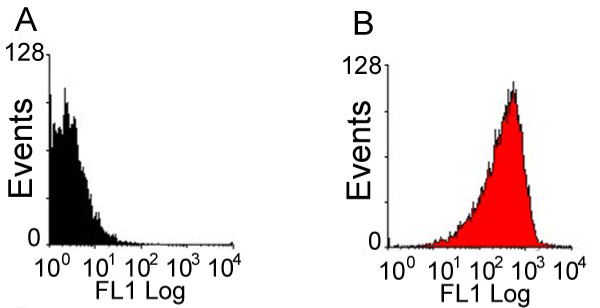
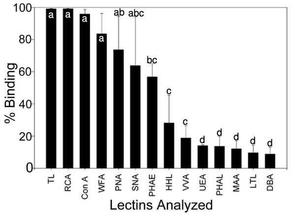
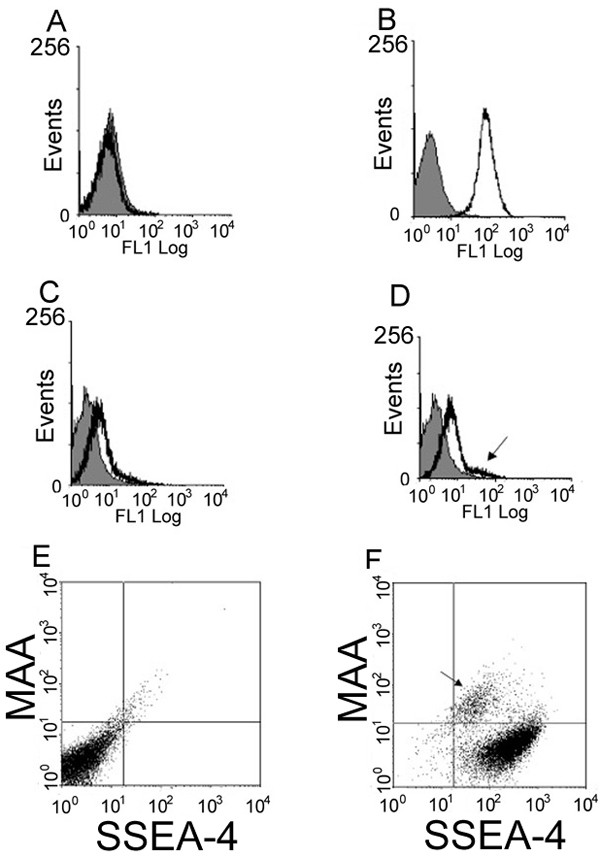
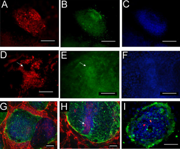
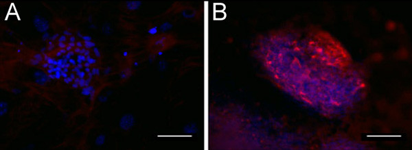
Results: Enriching cells for SSEA-4 expression increased the percentage of SSEA-4 positive cells to 98-99%. Using enriched high SSEA-4-expressing hESCs, we then analyzed the binding percentages of selected lectins and found a large variation in binding percentages ranging from 4% to 99% binding. Lycopersicon (tomato)esculetum lectin (TL), Ricinus communis agglutinin (RCA), and Concanavalin A (Con A) bound to SSEA-4 positive regions of hESCs and with similar binding percentages as SSEA-4. In contrast, we found Dolichos biflorus agglutinin (DBA) and Lotus tetragonolobus lectin (LTL) did not bind to hESCs while Phaseolus vulgaris leuco-agglutinin (PHA-L), Vicia villosa agglutinin (VVA), Ulex europaeus agglutinin (UEA), Phaseolus vulgaris erythro-agglutinin (PHA-E), and Maackia amurensis agglutinin (MAA) bound partially to hESCs. These binding percentages correlated well with immunocytochemistry results.

Conclusion: Our results provide information about types of carbohydrates and carbohydrate linkages found on pluripotent hESC surfaces. We propose that TL, RCA and Con A may be used as markers that are associated with the pluripotent state of hESCs because binding percentages and binding localization of these lectins are similar to those of SSEA-4. Non-binding lectins, DBA and LTL, may identify differentiated cell types; however, we did not find these lectins to bind to pluripotent SSEA-4 positive hESCs. This work represents a fundamental base to systematically classify pluripotent hESCs, and in future studies these lectins may be used to distinguish differentiated hESC types based on glycan presentation that accompanies differentiation.


Figures

Figure 1
Figure 1
Structure of Stage Specific Embryonic Antigen 4 (SSEA4). SSEA-4, one of the most commonly used hESC surface pluripotency markers, is a globoseries glycolipid that is characteristically downregulated upon hESC differentiation.
Figure 2
Figure 2
SSEA-4 expression in enriched hESCs determined by flow cytometry. The histogram plots of unstained, control hESCs (2A) and cells stained with SSEA-4 antibody after enrichment using magnetic bead sorting at day 0 (2B).
Figure 3
Figure 3
Quantitation of lectin binding on pluripotent hESC surfaces with 14 different lectins. The percent of cells with specific carbohydrate expression as determined by flow cytometry using 14 different lectins. The data are means +/- SD of 3 independent assays of BG01 and BG02 hESC lines. hESCs from each line were stained with one of 14 lectins and SSEA-4 immediately following enrichment for SSEA-4 expression. abcd: Means with different letters are significantly different, p < 0.05.
Figure 4
Figure 4
Flow cytometry histograms of lectin binding in unstained and stained HESCs. A-D shows histograms of SSEA-4 binding and representative lectins that were used in this study. To validate that double staining can be performed without signal interference, we determined that SSEA-4 expression was not found in the FL1 channel. Figure 4A shows a histogram plot with the overlay image of SSEA4 (black tracing) matching the histogram plot of unstained cells (grey fill). Figure 4B shows a positive peak shift in the histogram overlay with Tomato lectin (TL) in black tracing and unstained cells (grey fill). Figure 4C shows lack of Lotus tetragonolobus lectin (LTL) (black tracing) binding in the histogram overlay with unstained cells (grey fill). Figure 4D shows a histogram overlay with MAA (black tracing) binding and shows two peaks -one representing a large population that overlays unstained cells and a smaller population denoted by arrow that shows a smaller population of MAA+/SSEA4+ cells. Figure 4E shows plots of unstained hESCs, and Figure 4F shows subpopulations of cells characterized as SSEA-4+/MAA- or as SSEA-4+/MAA+ cells (arrow).
Figure 5
Figure 5
Carbohydrate expression as determined by lectin binding using immunocytochemistry. Figure 5A–C shows a hESC colony that represents uniform lectin binding. Ricinus Communis agglutinin (RCA) binding in red (5A) is shown throughout this SSEA-4 positive colony in green (5B). The DAPI nuclear stain image (blue) is also shown (5C). Other lectins showed partial binding patterns, such as Vicia Villosa agglutinin (VVA) binding (red), which is shown in a hESC colony (5D) that has uniform SSEA-4 antibody binding (green) (5E). Arrows denote distinct SSEA-4 positive regions lacking VVA binding. DAPI nuclear staining (blue) is also shown (5F). PHA-E binding is shown in two separate images in Figure 5G–H. In 5G there are two adjacent colonies, one that expresses strong binding of SSEA-4 antibody (green) and weak to no binding of PHA-E (red), and an adjacent colony showing binding of PHA-E without SSEA-4 antibody binding. (DAPI nuclear staining in blue). 5H, shows another colony with a streak of stacked cells (as determined by high DAPI expression, see arrow) in the middle of the colony that are beginning to lose SSEA-4 expression (green), but have strong PHA-E binding (red). However, the rest of the colony adjacent to this streak of cells is uniformly positive for SSEA-4 but is lacking PHA-E binding. 5I shows lack of DBA binding and presence of SSEA-4 and DAPI staining. Images and scale bars: 5A-G) 20× magnification, 100 μm. H-I) 10× magnification, 100 μm.
Figure 6
Figure 6
Addition of competitive sugar inhibitor blocks lectin expression as determined by immunocytochemistry. Addition of 200 mM galactose can block RCA binding in a hESC colony as shown by lack of RCA binding (6A), but does not affect DAPI stain (blue). However, in the absence of galactose, uniform binding of RCA (red) was detected 6(B). 6A-B) 20× magnification, 100 μm

Similar articles

Cited by

References

    1. Thomson JA, Itskovitz-Eldor J, Shapiro SS, Waknitz MA, Swiergiel JJ, Marshall VS, Jones JM. Embryonic stem cell lines derived from human blastocysts. Science. 1998;282:1145–1147. doi: 10.1126/science.282.5391.1145. - DOI - PubMed
    1. Shevinsky LH, Knowles BB, Damjanov I, Solter D. Monoclonal antibody to murine embryos defines a stage-specific embryonic antigen expressed on mouse embryos and human teratocarcinoma cells. Cell. 1982;30:697–705. doi: 10.1016/0092-8674(82)90274-4. - DOI - PubMed
    1. Kannagi R, Cochran NA, Ishigami F, Hakomori S, Andrews PW, Knowles BB, Solter D. Stage-specific embryonic antigens (SSEA-3 and -4) are epitopes of a unique globo-series ganglioside isolated from human teratocarcinoma cells. Embo J. 1983;2:2355–2361. - PMC - PubMed
    1. Henderson JK, Draper JS, Baillie HS, Fishel S, Thomson JA, Moore H, Andrews PW. Preimplantation human embryos and embryonic stem cells show comparable expression of stage-specific embryonic antigens. Stem Cells. 2002;20:329–337. doi: 10.1634/stemcells.20-4-329. - DOI - PubMed
    1. Draper JS, Pigott C, Thomson JA, Andrews PW. Surface antigens of human embryonic stem cells: changes upon differentiation in culture. J Anat. 2002;200:249–258. doi: 10.1046/j.1469-7580.2002.00030.x. - DOI - PMC - PubMed

LinkOut - more resources