. 2005 Jul 20;25(29):6755-64.
doi: 10.1523/JNEUROSCI.1247-05.2005.

Conditional inactivation of presenilin 1 prevents amyloid accumulation and temporarily rescues contextual and spatial working memory impairments in amyloid precursor protein transgenic mice

Affiliations

Conditional inactivation of presenilin 1 prevents amyloid accumulation and temporarily rescues contextual and spatial working memory impairments in amyloid precursor protein transgenic mice

Carlos A Saura et al. J Neurosci. .

Abstract

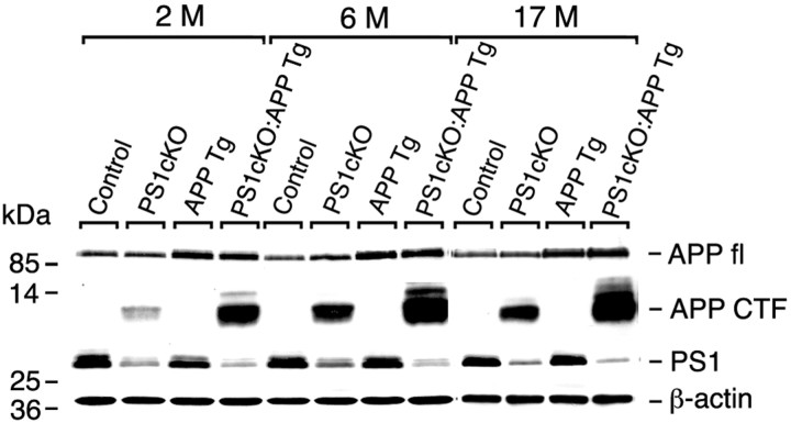
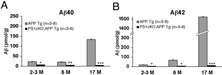
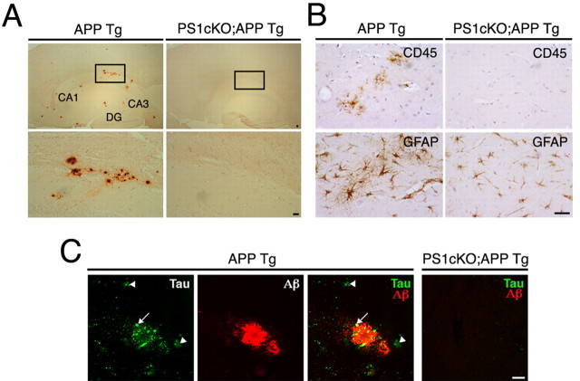
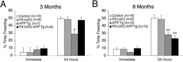
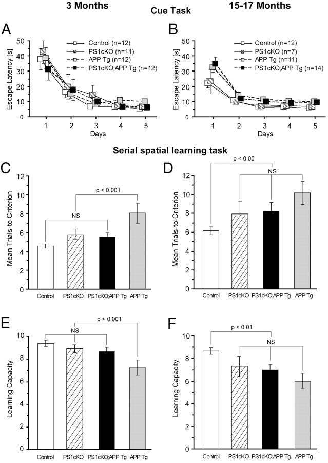
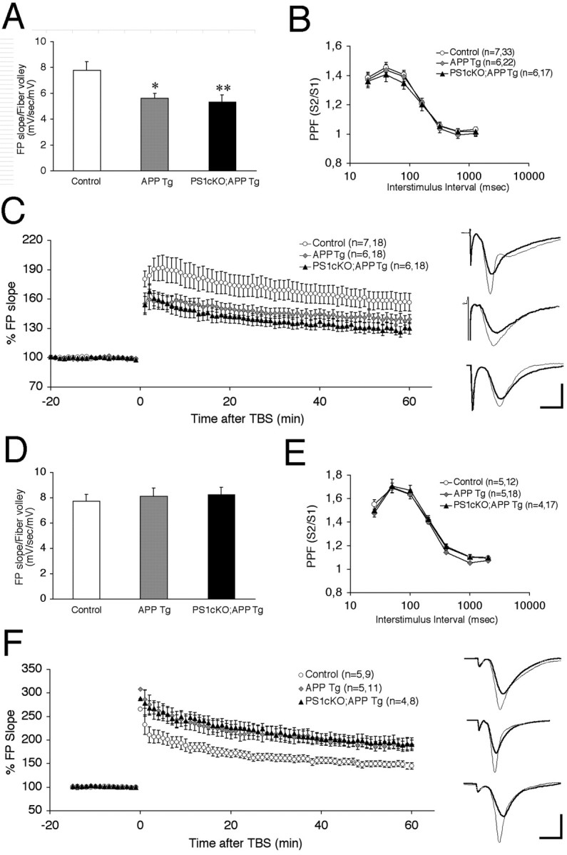
Accumulation of beta-amyloid (Abeta) peptides in the cerebral cortex is considered a key event in the pathogenesis of Alzheimer's disease (AD). Presenilin 1 (PS1) plays an essential role in the gamma-secretase cleavage of the amyloid precursor protein (APP) and the generation of Abeta peptides. Reduction of Abeta generation via the inhibition of gamma-secretase activity, therefore, has been proposed as a therapeutic approach for AD. In this study, we examined whether genetic inactivation of PS1 in postnatal forebrain-restricted conditional knock-out (PS1 cKO) mice can prevent the accumulation of Abeta peptides and ameliorate cognitive deficits exhibited by an amyloid mouse model that overexpresses human mutant APP. We found that conditional inactivation of PS1 in APP transgenic mice (PS1 cKO;APP Tg) effectively prevented the accumulation of Abeta peptides and formation of amyloid plaques and inflammatory responses, although it also caused an age-related accumulation of C-terminal fragments of APP. Short-term PS1 inactivation in young PS1 cKO;APP Tg mice rescued deficits in contextual fear conditioning and serial spatial reversal learning in a water maze, which were associated with APP Tg mice. Longer-term PS1 inactivation in older PS1 cKO;APP Tg mice, however, failed to rescue the contextual memory and hippocampal synaptic deficits and had a decreasing ameliorative effect on the spatial memory impairment. These results reveal that in vivo reduction of Abeta via the inactivation of PS1 effectively prevents amyloid-associated neuropathological changes and can, but only temporarily, improve cognitive impairments in APP transgenic mice.


Figures

Figure 1.
Figure 1.
Age-dependent accumulation of APP C-TFs in PS1 cKO;APP Tg mice. Levels of APP, APP C-TFs, and PS1 in cortical lysates of control, PS1 cKO, APP Tg, and PS1 cKO;APP Tg mice at 2, 6, and 17 months of age were analyzed by Western blotting with the use of antibodies specific for the C terminal of APP (Saeko) or PS1. An antibody specific for β-actin was used as a loading control. Levels of APP C-TFs in PS1 cKO;APP Tg mice were increased at 6 and 17 months of age compared with 2 months of age.
Figure 2.
Figure 2.
Accumulation of APPC-TFs in presynaptic terminals of PS1 cKO;APP Tg mice. A, Confocal microscopic images showing a marked increase in APP C-TFs recognized by C7 immunoreactivity (green) and a lack of colocalization (yellow) between the C7 immunoreactivity and the somatodendritic marker MAP2 immunoreactivity (red) in CA1 pyramidal neurons of 6-month-old PS1 cKO;APP Tg mice. B, Confocal microscopic images showing an increase in APP C-TFs recognized by C7 immunoreactivity (green) and its partial colocalization (yellow) with the postsynaptic marker NMDAR1 immunoreactivity (NR1; red) in CA1 pyramidal neurons of PS1 cKO;APP Tg mice. C, Confocal microscopic images showing a marked increase in APP C-TFs recognized by C7 immunoreactivity (green) and its abundant colocalization (yellow) with the presynaptic marker synaptophysin immunoreactivity (Syn; red) in CA1 pyramidal neurons of PS1 cKO;APP Tg mice. D, Electron microscopic images showing higher levels of accumulation of APP C-TFs in presynaptic compartments (arrowheads) compared with postsynaptic compartments (asterisk) in CA1 (top) and cortical (bottom) neurons from PS1 cKO;APP Tg mice at 6 months of age. Magnification: top, 19,000×; bottom, 13,500×. Scale bars: A-C, 25 μm; D, 500 nm.
Figure 3.
Figure 3.
Reduced accumulation of Aβ peptides in PS1 cKO;APP Tg mice. Levels of Aβ40 (A) and Aβ42 (B) in cortical lysates of APP Tg and PS1 cKO;APP Tg mice were quantified by ELISA. In APP Tg mice, the levels of Aβ40 and Aβ42 were increased from 2 to 17 months of age; levels of Aβ42 were higher than those of Aβ40 at 6 and 17 months of age. In the PS1 cKO;APP Tg mice, Aβ40 and Aβ42 were reduced significantly at all ages. Data represent the mean ± SEM; pmol/g signifies picomoles of Aβ per gram of cortex. *p < 0.01; **p < 0.001; ***p < 0.0001.
Figure 4.
Figure 4.
Absence of amyloid plaques and inflammation in PS1 cKO;APP Tg mice. A, Sagittal brain sections from APP Tg and PS1 cKO;APP Tg mice were stained with an Aβ antibody (R1282) to reveal the presence of amyloid plaques in the hippocampus of APP Tg mice and their absence in PS1 cKO;APP Tg mice at 6 months of age. Higher-power views of the boxed areas in the top panels are shown at the bottom. CA1 and CA3, CA1 and CA3 fields of the hippocampus, respectively; DG, dentate gyrus. Scale bar, 10 μm. B, Brain sections of APP Tg and PS1 cKO;APP Tg mice at 6 months of age were stained with antibodies specific for CD45 and GFAP. Activated microglial cells labeled by CD45 antibody and reactive astrocytes, which express high levels of GFAP and extend elaborate processes, are found only in the hippocampus of APP Tg mice, but not in PS1 cKO;APP Tg mice. Scale bar, 10 μm. C, Confocal microscopy analysis of APP Tg and PS1 cKO;APP Tg brain sections double-labeled with antibodies for phosphorylated tau (green) and Aβ (red). Dystrophic axons immunoreactive for phosphorylated tau either extend into plaques (arrow) or remain in the surrounding area of the plaque (arrowheads) in the hippocampus of APP Tg mice, whereas the PS1 cKO;APP Tg brain lacks such processes. Scale bar, 10 μm.
Figure 5.
Figure 5.
Age-dependent rescue of contextual memory in PS1 cKO;APP Tg mice. A, Control (n = 10), PS1 cKO (n = 9), APP Tg (n = 7), and PS1 cKO;APP Tg (n = 9) mice at 3 months of age were tested in the one-shock contextual fear conditioning task. All four groups showed similar levels of freezing immediately after the footshock. Control, PS1 cKO, and PS1 cKO;APP Tg mice showed similar levels of freezing (∼50%) at 24 h, whereas APP Tg mice exhibited significantly reduced levels of freezing (∼30%) (F(3,31) = 3.39; *p < 0.03). B, Control (n = 16), PS1 cKO (n = 6), APP Tg (n = 12), and PS1 cKO;APP Tg (n = 13) mice at 6 months of age were tested in the one-shock contextual fear conditioning task. All four groups showed similar levels of freezing immediately after the footshock. However, APP Tg and PS1 cKO;APP Tg mice showed reduced levels of freezing when compared with control and PS1 cKO mice at 24 h (F(3,43) = 9.0; **p < 0.0001). Data represent the mean ± SEM.
Figure 6.
Figure 6.
Age-dependent rescue of serial spatial learning in PS1 cKO;APP Tg mice. A, B, Control, PS1 cKO, APP Tg, and PS1 cKO;APP Tg mice at 3 months of age (A) and 15-17 months of age (B) were trained in the visible platform version of the water maze for 5 d consecutively (4 trials/d). APP Tg and PS1 cKO;APP Tg mice showed normal performance in the cue task. The number of mice used in each group is indicated in parentheses. C, Mice were trained in the serial spatial reversal task in a water maze for 10 d. Analysis of trials to reach criterion revealed no difference among control, PS1 cKO, and PS1 cKO;APP Tg groups (p > 0.10), but these three groups took fewer trials to learn the five platform locations than the APP Tg group (p < 0.001). These data indicate that PS1 cKO;APP Tg mice at 3 months of age exhibit a rescue of spatial working memory. D, Analysis of trials-to-criterion of mice at 15-17 months of age indicated that performances of the PS1 cKO, APP Tg, and PS1 cKO;APP Tg mice did not differ from each other (p > 0.10), and their performances were poorer than that shown by the control group (p < 0.05). E, Learning capacity was measured as the total number of spatial reversal locations learned in 10 d. Analysis of learning capacity showed similar learning capacity for the control, PS1 cKO, and PS1 cKO;APP Tg groups and reduced learning capacity in the APP Tg group at 3 months of age (p < 0.001). These data indicate a rescue of learning capacity in PS1 cKO;APP Tg mice at 3 months of age. F, The learning capacity of APP Tg, PS1 cKO;APP Tg, and PS1 cKO groups at 15-17 months of age did not differ significantly, but the average performance of these three groups was significantly lower than that of the control group (p < 0.01). Data represent the mean ± SEM.
Figure 7.
Figure 7.
Similar hippocampal synaptic alterations in APP Tg and PS1 cKO;APP Tg mice. A, Basal synaptic transmission is reduced in APP Tg and PS1 cKO;APP Tg mice at 6 months of age. The I/O slope was obtained by plotting the FV amplitude against the initial slope of the evoked fEPSP in acute hippocampal slices of control, APP Tg, and PS1 cKO;APP Tg mice. The graph represents the average slope of the I/O curves. *p < 0.02; **p < 0.005. B, The graph depicts the paired pulse response ratio (second fEPSP/first fEPSP) obtained at different interstimulus intervals in 6-month-old mice. APP Tg and PS1 cKO;APP Tg mice show normal PPF (p = 0.14). C, Left, Time course of the effects of five episodes of TBS on the fEPSP initial slope in 6-month-old mice. Right, Examples of LTP induced in slices from control (top), APP Tg (middle), and PS1 cKO;APP Tg (bottom) mice. Super imposed traces are averages of four consecutive responses recorded 1 min before (thin line) and 60 min after (thick line) TBS. APP Tg and PS1 cKO;APP Tg mice show impaired LTP. D, The graph represents the average slope of the I/O curves in 3-month-old mice. The average of FV-fEPSP slopes in APP Tg and PS1 cKO;APP Tg mice is similar to that of control mice at 3 months of age (p > 0.05). E, The graph depicts the paired pulse response ratio obtained at different interstimulus intervals in 3-month-old mice. APP Tg, PS1 cKO;APP Tg, and control mice show similar PPF (p = 0.13). F, Left, Time course of the effects of five episodes of TBS on the fEPSP initial slope in 3-month-old mice. Right, Examples of LTP induced in slices from control (top), APP Tg (middle), and PS1 cKO;APP Tg (bottom) mice. Super imposed traces are averages of four consecutive responses recorded 1 min before (thin line) and 60 min after TBS (thick line). In B, C, E, F, the numbers of mice (left) and slices (right) are indicated in parentheses. Data represent the mean ± SEM. Calibration: (in C, F) 0.5 mV, 5 ms.

Similar articles

Cited by

References

    1. Amtul Z, Lewis PA, Piper S, Crook R, Baker M, Findlay K, Singleton A, Hogg M, Younkin L, Younkin SG, Hardy J, Hutton M, Boeve BF, Tang-Wai D, Golde TE (2002) A presenilin 1 mutation associated with familial frontotemporal dementia inhibits γ-secretase cleavage of APP and Notch. Neurobiol Dis 9: 269-273. - PubMed
    1. Borchelt DR, Thinakaran G, Eckman CB, Lee MK, Davenport F, Ratovitsky T, Prada C-M, Kim G, Seekins S, Yager D, Slunt HH, Wang R, Seeger M, Levey AI, Gandy SE, Copeland NG, Jenkins NA, Price DL, Younkin SG, Sisodia SS (1996) Familial Alzheimer's disease-linked presenilin 1 variants elevate Aβ1-42/1-40 ratio in vitro and in vivo Neuron 17: 1005-1013. - PubMed
    1. Buxbaum J, Thinakaran G, Koliatsos V, O'Callahan J, Slunt H, Price D, Sisodia S (1998) Alzheimer amyloid protein precursor in the rat hippocampus: transport and processing through the perforant path. J Neurosci 18: 9629-9637. - PMC - PubMed
    1. Cai D, Leem JY, Greenfield JP, Wang P, Kim BS, Wang R, Lopes KO, Kim SH, Zheng H, Greengard P, Sisodia SS, Thinakaran G, Xu H (2003) Presenilin-1 regulates intracellular trafficking and cell surface delivery of β-amyloid precursor protein. J Biol Chem 278: 3446-3454. - PubMed
    1. Chapman P, White G, Jones M, Cooper-Blacketer D, Marshall V, Irizarry M, Younkin L, Good M, Bliss T, Hyman B, Younkin S, Hsiao K (1999) Impaired synaptic plasticity and learning in aged amyloid precursor protein transgenic mice. Nat Neurosci 2: 271-276. - PubMed

Publication types

MeSH terms