. 2005 Jul 20;25(29):6765-74.
doi: 10.1523/JNEUROSCI.1700-05.2005.

FasL (CD95L/APO-1L) resistance of neurons mediated by phosphatidylinositol 3-kinase-Akt/protein kinase B-dependent expression of lifeguard/neuronal membrane protein 35

Affiliations

FasL (CD95L/APO-1L) resistance of neurons mediated by phosphatidylinositol 3-kinase-Akt/protein kinase B-dependent expression of lifeguard/neuronal membrane protein 35

Christoph P Beier et al. J Neurosci. .

Abstract

The contribution of Fas (CD95/APO-1) to cell death mechanisms of differentiated neurons is controversially discussed. Rat cerebellar granule neurons (CGNs) express high levels of Fas in vitro but are resistant to FasL (CD95L/APO-1L/CD178)-induced apoptosis. We here show that this resistance was mediated by a phosphatidylinositol 3-kinase (PI 3-kinase)-Akt/protein kinase B (PKB)-dependent expression of lifeguard (LFG)/neuronal membrane protein 35. Reduction of endogenous LFG expression by antisense oligonucleotides or small interfering RNA lead to increased sensitivity of CGNs to FasL-induced cell death and caspase-8 cleavage. The inhibition of PI 3-kinase activity sensitized CGNs to FasL-induced caspase-8 and caspase-3 processing and caspase-dependent fodrin cleavage. Pharmacological inhibition of PI 3-kinase, overexpression of the inhibitory protein IkappaB, or cotransfection of an LFG reporter plasmid with dominant-negative Akt/PKB inhibited LFG reporter activity, whereas overexpression of constitutively active Akt/PKB increased LFG reporter activity. Overexpression of LFG in CGNs interfered with the sensitization to FasL by PI 3-kinase inhibitors. In contrast to CGNs, 12 glioma cell lines, which are sensitive to FasL, did not express LFG. Gene transfer of LFG into these FasL-susceptible glioma cells protected against FasL-induced apoptosis. These results demonstrate that LFG mediated the FasL resistance of CGNs and that, under certain circumstances, e.g., inhibition of the PI 3-kinase-Akt/PKB pathway, CGNs were sensitized to FasL.


Figures
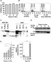
Figure 1.
Figure 1.
Resistance of CGNs against FasL-induced apoptosis and expression of LFG. A, The resistance of CGNs cultured in medium containing 10% FCS to 500 U/ml FasL did not change in the course of in vitro maturation. CGNs were resistant to 500 U/ml FasL at all time points (n = 3 per time point). B, To test the cross-reactivity of human FasL used in the experiments with rat Fas, rat lymphocytes isolated from spleens of 8-d-old rats were coincubated with FasL. Cell death was detected as the sub-G0/G1 peak by propidium iodide staining and flow cytometry at 16 h. C, D, The temporal profile of FLIP (long), FLIP (short), and LFG expression in CGNs was studied by Western blot analysis. Lysates from CGNs were prepared at the time points indicated, and 30 μg of protein was used per lane. As positive control lysates of HEK-293, cells were transfected with FLIP (long) and FLIP (short) using Effectene. FLIP was expressed in CGNs until DIV 5 but was no longer detected at DIV 8. In contrast, expression of LFG was upregulated from DIV 2 to DIV 8. E, LFG mRNA expression in CGNs in vitro (left) and in the cerebellum in vivo (right) during maturation. LFG mRNA expression was assessed using RT-PCR and compared with GAPDH expression (in vitro) or actin expression (in vivo) shown as relative data (n = 3 per time point; *p < 0.05, **p < 0.001, ANOVA followed by Scheffe's post hoc test.).
Figure 2.
Figure 2.
Colocalization of LFG and Fas in CGNs. A, CGNs were stained for LFG (green) and NeuN (red). All LFG-positive cells showed colocalization with NeuN (yellow). B, In contrast to all glioma cell lines tested, CGNs expressed LFG mRNA, as shown by Northern blot analysis. Methylene blue staining was performed as a loading control. C, Confocal image of Fas (red)-labeled and LFG (green)-labeled CGNs viewed in three dimensions after orthogonal reconstruction of z-scans with a thickness of 0.77 μm. There is colocalization of both stainings in neurites, cytoplasm, and the cell membrane. D, E, Fluorescein-labeled antisense oligonucleotides (green) efficiently transfected CGN. Antisense oligonucleotides to LFG reduced the LFG levels in CGN. After transfection, the uptake of oligonucleotides was reflected by green fluorescence. Cells were fixed and stained for LFG (red). Those neurons with a strong uptake of fluorescein-labeled oligonucleotides showed no or weak LFG immunostaining (blue arrows), whereas CGNs with a limited uptake were still positive for LFG (white arrows). F, LFG mRNA expression in CGNs, which were not transfected or transfected with antisense or scrambled oligonucleotides (oligo) for 18 h. LFG mRNA expression was assessed using RT-PCR and compared with actin expression shown as relative data (n = 3 per time point; *p < 0.05).
Figure 3.
Figure 3.
LFG in FasL-induced apoptosis. A, B, Incubation with antisense LFG (0.5 pm) or FasL (500 U/ml) for 24 h alone did not alter cell survival, whereas cotreatment resulted in cell death and caspase-8 cleavage. Exposure to scrambled oligonucleotide (oligo) alone or in combination with FasL had no such effect. Cell viability was assessed by FDA staining (n = 3 per condition; *p < 0.001, ANOVA followed by Scheffe's post hoc test). C, To analyze the efficacy of the pSUPER-LFG siRNA vector, HEK-293 cells were cotransfected with pcDNA3-LFG and pSUPER-LFG, pSUPER control, or the empty vector using FuGene. Cells were lysed at 48 h after transfection. D, The siRNA expressed in pSUPER-LFG sensitized CGNs to FasL-induced cell death in transfected cells. CaCl2 was used for transfection. Four hours after transient cotransfection of CGNs with GFP and pSUPER (ratio of 1:2), 450 cells were evaluated per condition (-FasL, +FasL). Another 12 and 24 h later, the survival of the transfected CGNs was assessed by counting again the same visual fields. The viability compared with untreated conditions is given [n = 3 per condition; *p < 0.05, **p < 0.01 compared with untreated (0 h) condition]. E, Stable overexpression of LFG protected LN-18 glioma cells from FasL-induced death. Viability was assessed by crystal violet staining (n = 3 per time point and condition). F, LN-18 glioma cells were protected from FasL-induced cell death when transiently cotransfected with GFP and LFG (GFP/LFG of 2:1). Cell death was assessed 18 h after addition of 50 U/ml FasL by propidium iodide staining, followed by flow cytometric analysis of DNA content in GFP-positive cells.
Figure 4.
Figure 4.
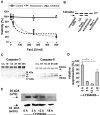
Inhibition of PI 3-kinase establishes sensitivity of CGNs to FasL. A-C, CGNS treated with wortmannin (0.2 μm), LY294002 (10 μm), or FasL (25-500 U/ml) alone for 24 h did not undergo apoptosis. Only the combination of wortmannin (0.2 μm) and FasL (500 U/ml) caused a significant decrease in cell viability as well as fodrin cleavage at 24 h and caspase-8 and caspase-3 cleavage at the time points indicated. For Western blot analysis, 30 μg of protein per lane was used (n = 3 per condition). D, E, CGNs treated with LY294002 (10 μm) downregulated LFG mRNA and protein expression. mRNA content was assessed using real-time-PCR (n = 2 per time point). LFG mRNA bands were normalized to 18 S mRNA expression and shown as relative data (*p < 0.05; ANOVA followed by Scheffe's post hoc test). Protein levels were assessed by Western blot analysis. LK, Low potassium.
Figure 5.
Figure 5.
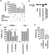
Activation of caspase-8 in FasL induced apoptosis of CGNs. A, B, The specific caspase-8 inhibitor IETD-fmk (100 μm) prevented cell death and fodrin cleavage induced by the combination of FasL (500 U/ml) and wortmannin (0.2 μm). Cell viability of CGNs was measured by FDA staining after 24 h of treatment with substances as indicated (n = 3 per condition; *p < 0.05, ANOVA followed by Scheffe's post hoc test). LK, Low potassium. C, CGNs from lpr mice were protected against cell death induced by the combination of the Fas agonistic antibody Jo2 (20 μg/ml) with wortmannin (0.2 μm) or LY294002 (10 μm), whereas CGNs obtained from strain-specific controls underwent apoptosis (n = 3 per condition; **p < 0.01, ANOVA followed by Scheffe's post hoc test). D, CGNs were cotransfected with GFP and pcDNA3-LFG or an empty vector (ratio 2:1) using the GeneGun. With this method ∼2-5% of cells were transfected. Approximately 300 GFP-positive cells in two wells per condition were evaluated per condition (LFG vs empty vector). CGNs were then treated with the combination of 500 U/ml FasL and 0.2 μm wortmannin. Another 24 h later, the survival of the transfected CGNs was assessed by counting again the same visual fields. The viability compared with pretreatment conditions is given (*p < 0.05; ANOVA followed by Scheffe's post hoc test).
Figure 6.
Figure 6.
LFG promoter activity regulated by Akt/PKB and NFκB in PC12 cells. A, PC12 cells were cotransfected (ratio of 4:1) with LFG-Luc and pCMV-RL and incubated as indicated (100 ng/ml NGF, 10 μm LY294002). Reporter gene activities were assessed after 16 h (n = 5; #p < 0.05, *p < 0.01, **p < 0.001, ANOVA followed by Scheffe's post hoc test). B, C, Cells were cotransfected with LFG-Luc, pCMV-RL, and the vectors given (ratio 4:1:1). Reporter gene activities were measured at 48 h after transfection and 16 h after treatment.

References

    1. Bechmann I, Mor G, Nilsen J, Eliza M, Nitsch R, Naftolin F (1999) FasL (CD95L, Apo1L) is expressed in the normal rat and human brain: evidence for the existence of an immunological brain barrier. Glia 27: 62-74. - PubMed
    1. Bellacosa A, Testa JR, Staal SP, Tsichlis PN (1991) A retroviral oncogene, akt, encoding a serine-threonine kinase containing an SH2-like region. Science 254: 274-277. - PubMed
    1. Brummelkamp TR, Bernards R, Agami R (2002) A system for stable expression of short interfering RNAs in mammalian cells. Science 296: 550-553. - PubMed
    1. Brunet A, Bonni A, Zigmond MJ, Lin MZ, Juo P, Hu LS, Anderson MJ, Arden KC, Blenis J, Greenberg ME (1999) Akt promotes cell survival by phosphorylating and inhibiting a Forkhead transcription factor. Cell 96: 857-868. - PubMed
    1. Burgering BMT, Coffer PJ (1995) Protein kinase B (c-Akt) in phosphatidylinositol-3-OH kinase signal transduction. Nature 376: 599-602. - PubMed

Publication types

MeSH terms