. 2005 Jul 20;25(29):6816-25.
doi: 10.1523/JNEUROSCI.1114-05.2005.

Neonatal and adult neurogenesis provide two distinct populations of newborn neurons to the mouse olfactory bulb

Affiliations

Neonatal and adult neurogenesis provide two distinct populations of newborn neurons to the mouse olfactory bulb

Morgane Lemasson et al. J Neurosci. .

Abstract

In mammals, the olfactory bulb (OB) constitutes one of two regions of the postnatal brain with continuous neurogenesis throughout life. Despite intense explorations of neuronal replacement in the adult OB, little is known about the mechanisms that operate at earlier postnatal stages. This question is particularly pertinent, because the majority of local interneurons are born in the neonate, when olfaction controls vital functions. Here, we analyzed the recruitment of newborn cells to the granule cell (GC) layer (GCL) and found that the postnatal mouse OB is supplied with two spatiotemporally distinct populations of newborn interneurons. Early born [postnatal day 3 (P3) to P7] GCs constitute a threefold larger population compared with those generated later (P14-P60), and some of them are produced locally within the OB itself. Newborn interneurons generated at P3-P7 were predominantly targeted to the external edge of the GCL, whereas newly generated cells were positioned deeper in older mice. Additionally, although approximately 50% of adult newborn cells were eliminated within a few weeks of reaching the OB, almost the entire population of early born GCs survived until adulthood. Importantly, early olfactory experience specifically modifies the number of newborn GCs in neonates but leaves unaltered the amount of neurons generated during adulthood. Together, these results demonstrate that early postnatal neurogenesis endows the neonate bulbar circuit with newborn GCs that differ morphologically and functionally from those produced in the adult.


Figures
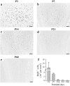
Figure 1.
Figure 1.
Local cellular proliferation in the postnatal olfactory bulb decreases with age. a-e, Representative images of BrdU+ cells located in the granule cell layer of 3-d-old (a), 7-d-old (b), 14-d-old (c), 21-d-old (d), and 60-d-old (e) mice. f, Mean density of BrdU+ cells (per mm2) in the granule cell layer determined 2 h after a single injection of BrdU (n = 2-4 animals per group). Scale bar, 50 μm.
Figure 2.
Figure 2.
Age-dependent changes in the migration duration required for neuroblasts to reach the OB. a, Representative images of a sagittal section of a 19-d-old mouse brain demonstrating the pattern of distribution of BrdU+ cells generated at P3 along the SVZ/OB pathway. LV, Lateral ventricle. b, Coronal sections of the OB of animals injected with BrdU at P3, P7, P14, P21, and P60 and killed following different survival periods as indicated in the parentheses. The coronal sections were taken at the level of the dotted line indicated in the sagittal section in a. Note the disappearance of BrdU+ cells in the RMSOB following the chosen survival times. c, Ratio of BrdU+ cells in the RMSOB to those in the OB. Scale bars: a, 500 μm; b, 50 μm.
Figure 3.
Figure 3.
Age-dependent decreases in the density of surviving newly generated cells in the granule cell layer. a, Mean number of BrdU+ cells (per mm2) in the granule cell layer at different ages. Animals (n = 3-5 per group) were killed following a time window required to empty the RMS (as indicated in Fig. 2). b, Mean number of BrdU+ cells (per mm2) throughout the rostrocaudal axis of the OB. c, Ratio of SVZ/RMS-generated cells in the OB after migration versus that of locally proliferating cells at different ages. Note that, because of the decreased local OB proliferation with age, this ratio increases during postnatal development.
Figure 4.
Figure 4.
Targeting of newborn granule cells changes during bulbar development. a, e, Quantifying BrdU+ cell distribution throughout the internal-external (a) and ventrodorsal (e) axes of the granule cell layer (see Materials and Methods for details). EPL, External plexiform layer; GL, glomerular layer. b, c, Quantification of BrdU+ cells throughout the internal-external axis expressed as percentage (b) and density (c). Note the different age-dependent distribution profiles of BrdU+ cells. d, Internal-external distribution of BrdU+ cells measured after short-term (2 h; black circles) or long-term (from 20 to 28 d, depending on postnatal age of animals; white circles) survival. f, g, Distribution of BrdU+ cells throughout the ventrodorsal axis expressed as percentage (f) and density (g). Note the equal distribution of BrdU+ profiles across ages. Scale bars, 200 μm.
Figure 5.
Figure 5.
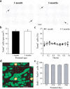
Cell death and fate of newborn cells remain constant in the olfactory bulb at different postnatal ages. a, TUNEL+ cells (arrows) in the middle of granule cell layer of 1-month-old (left) and 3-month-old (right) mice. b, Number of apoptotic nuclei (per mm2) in the granule cell layer of young and adult mice. c, Distribution of TUNEL+ cells (expressed as percentage) within the internal-external axis of the granule cell layer of 1- and 3-month-old animals (n = 4 per group). d, Example of a 3D-reconstructed BrdU+ cell (red) colabeled with the neuronal marker NeuN (green) in the granule cell layer. Reconstructed orthogonal projections are presented as viewed in the x-z (top) and y-z (right) planes. e, Percentage of double-labeled cells in the granule cell layer of 7-, 14-, 21-, and 60-d-old mice. More than 1000 cells, taken from three animals per group, were analyzed for each postnatal age. Scale bars: a, 20 μm; d, 10 μm. Error bars indicate SEM.
Figure 6.
Figure 6.
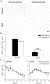
Different survival rates of newborn granule cells in the neonate and the adult olfactory bulb. a, Photomicrographs of BrdU-immunostained sections of the granule cell layer in early postnatal (top; P4) and adult mice (bottom; P45) 22 d (left) and 60 d (right) after BrdU injections. b, Corresponding densities of BrdU+ cells generated during different periods of life and allowed to survive for short (22 d) and long (60 d) BrdU postinjection times (n = 4-10 animals per group). Note the differences in age-dependent persistence of newborn cells in young and adult mice. c, Distribution of early- and late-generated GCs following different survival periods. Note the distribution differences between early postnatal (P4) and adult animals (P45) versus the similar distributions observed following different survival periods. **p < 0.001. Scale bar, 20 μm. Error bars indicate SEM.
Figure 7.
Figure 7.
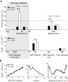
Early olfactory experience alters early but not late postnatal neurogenesis. a, Boxed area, Pups were reared in a citral- or saline-scented environment from E18 to P9. Top graph, P8 and P60 mice were subjected to a preference test. Histograms indicate the ratio of time spent by saline- and citral-reared animals (black and white bars, respectively) investigating citral or carvon shavings versus clean shavings (n = 9-33 animals per group). At both ages, animals show a significant preference to citral compared with saline. The specificity of this preference is shown by the absence of preference to carvon shavings at any age. Bottom graph, Quantification of BrdU+ cells following early olfactory experience. BrdU was administered either from P4 to P7 (one injection per day) or at P45 (four injections), and animals were killed for immunohistochemistry 20 d later. The mean number of newborn GCs was significantly higher in citral-reared animals compared with control mice when measured at P27 but not at P65. b, Spatial distribution of BrdU+ cells counted in the granule cell layer along the rostrocaudal axis (left panel), the internal-external axis (middle panel), and the ventrodorsal axis (right panel) of the OB taken from saline- or citral-reared mice. Animals were injected with BrdU from P4 to P7 (n = 5-7 animals per group). ***p < 0.001 with a Student's t test.

References

    1. Altman J (1969) Autoradiographic and histological studies of postnatal neurogenesis. IV. Cell proliferation and migration in the anterior forebrain, with special reference to persisting neurogenesis in the olfactory bulb. J Comp Neurol 137: 433-457. - PubMed
    1. Altman J, Das GD (1966) Autoradiographic and histological studies of postnatal neurogenesis. I. A longitudinal investigation of the kinetics, migration and transformation of cells incorporating tritiated thymidine in neonate rats, with special reference to postnatal neurogenesis in some brain regions. J Comp Neurol 126: 337-389. - PubMed
    1. Baker H, Liu N, Chun HS, Saino S, Berlin R, Volpe B, Son JH (2001) Phenotypic differentiation during migration of dopaminergic progenitor cells to the olfactory bulb. J Neurosci 21: 8505-8513. - PMC - PubMed
    1. Bayer SA (1983) 3H-Thymidine-radiographic studies of neurogenesis in the rat olfactory bulb. Exp Brain Res 50: 329-340. - PubMed
    1. Belluzzi O, Benedusi M, Ackman J, LoTurco JJ (2003) Electrophysiological differentiation of new neurons in the olfactory bulb. J Neurosci 23: 10411-10418. - PMC - PubMed

Publication types