. 2005 Jul 20;25(29):6845-56.
doi: 10.1523/JNEUROSCI.0442-05.2005.

Endocannabinoid signaling in rat somatosensory cortex: laminar differences and involvement of specific interneuron types

Affiliations

Endocannabinoid signaling in rat somatosensory cortex: laminar differences and involvement of specific interneuron types

Agnes L Bodor et al. J Neurosci. .

Abstract

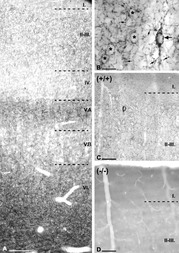
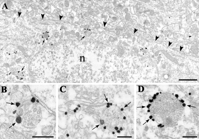
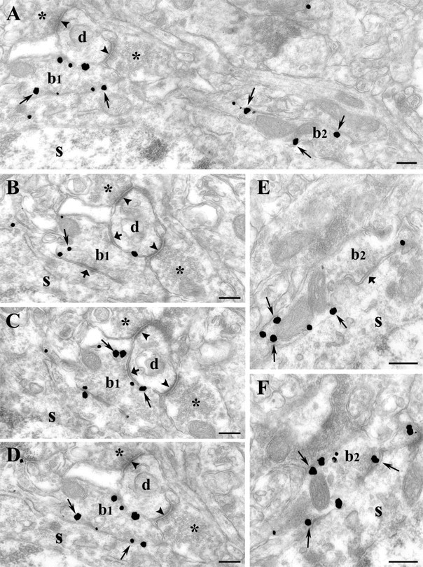
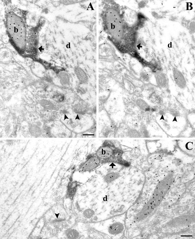
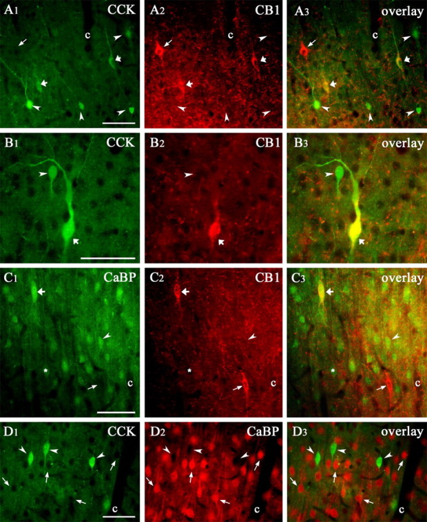
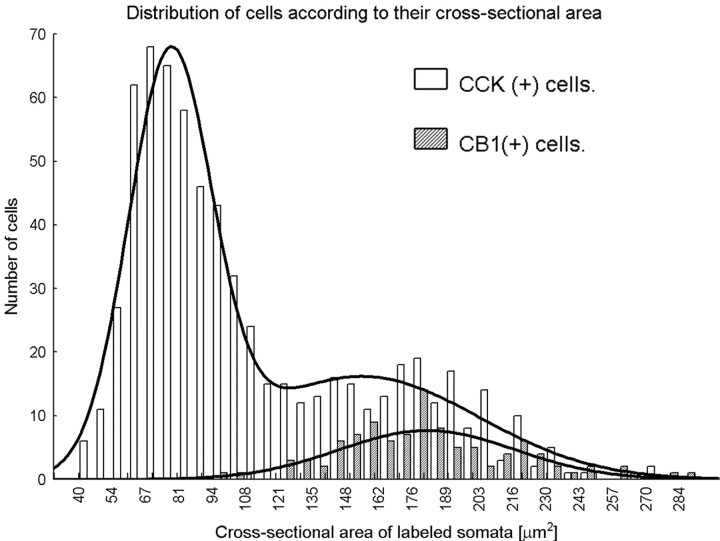
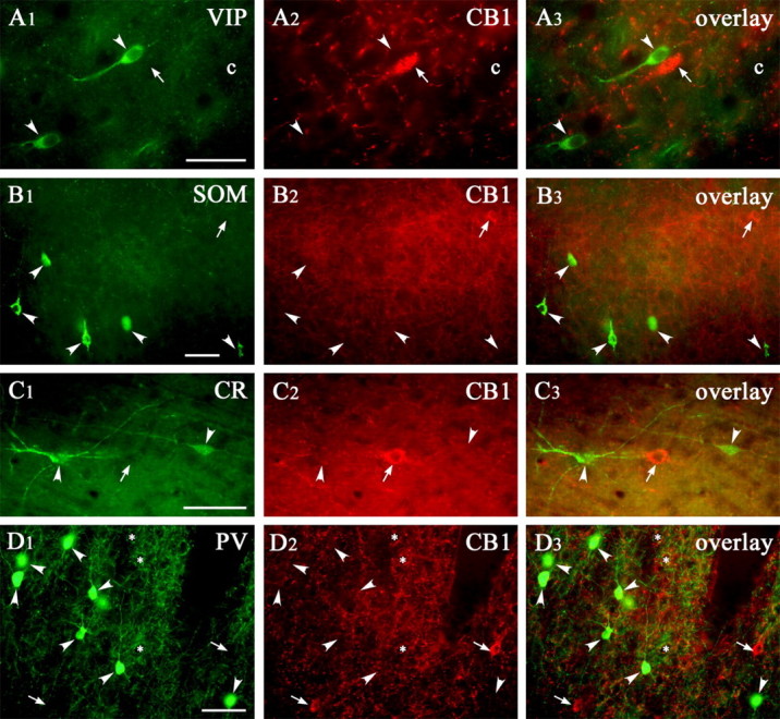
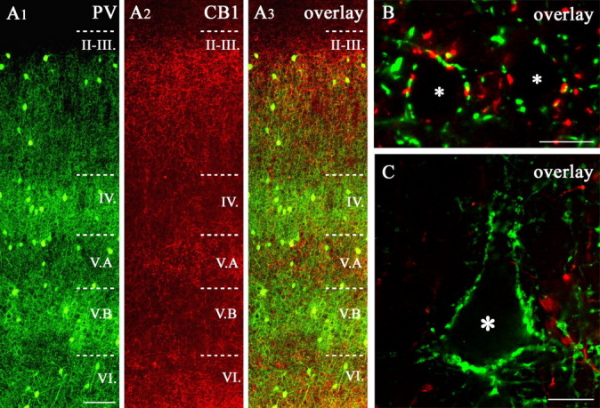
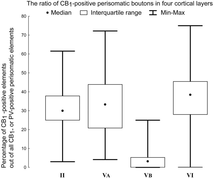
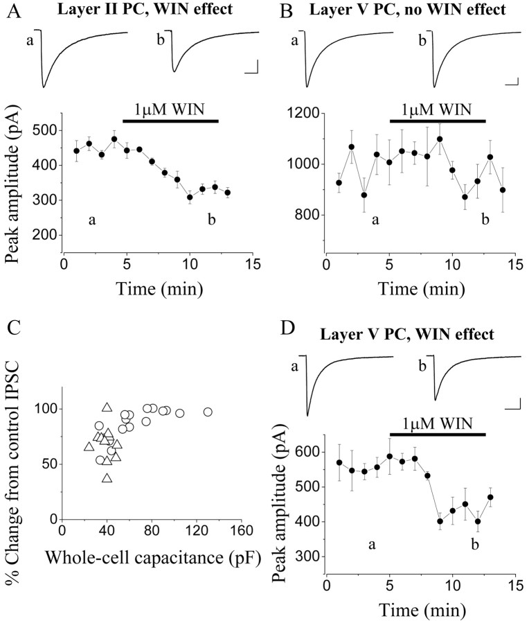
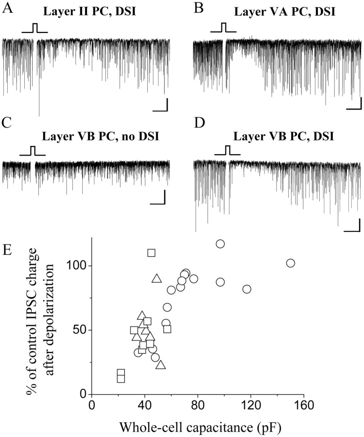
Endocannabinoid-mediated retrograde signaling exerts powerful control over synaptic transmission in many brain areas. However, in the neocortex, the precise laminar, cellular, and subcellular localization of the type 1 cannabinoid receptor (CB1) as well as its function has been elusive. Here we combined multiple immunolabeling with whole-cell recordings to investigate the morpho-functional characteristics of cannabinoid signaling in rat somatosensory cortex. Immunostaining for CB1 revealed axonal and somatic labeling with striking layer specificity: a high density of CB1-positive fibers was seen in layers II-III, in layer VI, and in upper layer V, whereas other layers had sparse (layer IV) or hardly any (layer I) staining. Membrane staining for CB1 was only found in axon terminals, all of which contained GABA and formed symmetric synapses. Double immunostaining also revealed that CB1-positive cells formed two neurochemically distinct subpopulations: two-thirds were cholecystokinin positive and one-third expressed calbindin, each subserving specific inhibitory functions in cortical networks. In addition, cannabinoid sensitivity of GABAergic input showed striking layer specificity, as revealed by both electrophysiological and anatomical experiments. We found a unique population of large pyramidal neurons in layer VB that received much less perisomatic innervation from CB1-expressing GABAergic axon terminals and, accordingly, showed no depolarization-induced suppression of inhibition, unlike pyramidal cells in layer II, and a population of small pyramidal cells in layer V. This suggests that inhibitory control of pyramidal cells involved in intracortical or corticostriatal processing is fine-tuned by activity-dependent endocannabinoid signaling, whereas inhibition of pyramidal cells relaying cortical information to lower subcortical effector centers often lacks this plasticity.


Figures

Figure 1.
Figure 1.
Distribution of CB1 cannabinoid receptors in the rat somatosensory cortex. A, Immunostaining of the rat somatosensory cortex for CB1 reveals axonal labeling in all layers with variable densities. The highest density of labeled fibers can be seen in layers VI, II-III, and VA. The apparently very high level of staining at the bottom of layer VI is primarily attributable to darker background of well contrasted myelinated fibers leaving the cortex. Analysis shows that this layer is labeled similarly to layer II (see also Fig. 8). Immunostained axons are sparse in layer VB and layer IV, whereas layer I has only negligible labeling (C). B, Both synaptic varicosities (small arrows) and preterminal, thin axon segments show strong CB1 immunostaining immunostaining, as seen in the higher-power micrograph of layers II-III. A large number of varicosities surround the CB1-negative somata of pyramidal cells (asterisks in B) in a basket-like manner. CB1-immunoreactive large cell bodies show the characteristics of interneurons (large arrows). C, CB1-immunopositive axons delineate the border between layer I and layers II-III in the cortex of wild-type mice, as in the rat. D, Immunostaining for CB1 receptors in CB1-/- mice results in a complete lack of labeling, confirming the selectivity of the antibody for CB1. Scale bars: A, C, D, 100 μm; B, 20 μm.
Figure 2.
Figure 2.
Compartmental distribution of CB1 cannabinoid receptors in the cell bodies of interneurons. A-D, CB1 cannabinoid receptors in cell bodies are restricted to intracellular membrane compartments. Note the lack of labeling on the plasma membrane (arrowheads in A). In contrast, immunogold particles (arrows in B-D), representing the localization of CB1 receptor protein, are frequently attached to elements of the endosome-lysosome system or multivesicular bodies (thin arrows in A). Some are also found on the Golgi complex and on the rough endoplasmic reticulum. n, Nucleus. B-D, The silver gold-particles (arrows), labeling the C terminus of the CB1 receptors, are always located on the outer surface of these organelles, suggesting a transport function to or from the plasma membrane of axons and a correct insertion (C terminus intracellular) of the receptor. Scale bars: A, 1 μm; B-D, 0.25 μm.
Figure 3.
Figure 3.
Presynaptic localization of CB1 on inhibitory axon terminals in rat somatosensory cortex. A-F, In layers II-III, silver-intensified gold particles (thin arrows) representing the localization of CB1-immunoreactivity are found on boutons (b1 and b2) but not on dendrites or somata. The antibody directed against the intracellular C-terminal portion of CB1 labels the intracellular side of the membrane of the terminals. B-D, Serial sections cut from b1 forming two symmetrical synapses (thick arrow), one on a soma (s) and the other on a dendritic shaft (d). This dendritic shaft also receives two asymmetrical synapses (arrowheads) from CB1-negative, presumably excitatory, axon terminals (asterisk). E, F, High-power electron micrographs show serial sections cut from a second bouton (b2). This axon terminal also forms a symmetrical synapse (thick arrow) on the same cell body (s) as b1 and is densely covered by gold particles (thin arrow). The CB1-immunoreactive axon terminals frequently contain dense-core vesicles (in D, in bouton 1). Scale bars: A-E, 0.25 μm.
Figure 4.
Figure 4.
Presynaptic localization of CB1 on GABA-positive axon terminals in rat somatosensory cortex. A-C, Electron micrographs of CB1-immunoreactive boutons (visualized by immunoperoxidase staining using the DAB precipitate) forming symmetrical synapses (thick arrows) on CB1-negative pyramidal cell dendrites (d, in A-C). Postembedding immunogold staining performed on these ultrathin sections demonstrates that all CB1-positive boutons are immunoreactive for GABA (see accumulation of colloidal gold particles over the mitochondria of DAB-labeled boutons; particles are accumulated over mitochondria attributable to the fixation method used). Axon terminals that form asymmetrical synapses (arrowhead) on dendritic spines were always negative for both GABA and CB1. These correspond to glutamatergic boutons and can be used to indicate the level of background staining for GABA. Scale bars: A-C, 0.2 μm.
Figure 5.
Figure 5.
CB1 receptors are expressed by two neurochemically distinct subpopulations of interneurons in the rat somatosensory cortex. A1-A3, B1-B3, CCK/CB1 double-labeled cells are shown by the thick arrows. On the overlay, they appear in yellow (CCK immunostaining in A1 and B1; CB1 immunostaining in A2 and B2; the overlay in A3 and B3). The arrowheads show cells that are positive only for CCK, and the thin arrow points to a CB1-positive cell devoid of CCK. Two CCK-positive cells are shown in B1, a large (thick arrow) and a small (arrowhead), but only the large one expresses CB1 (B2). C1-C3, CaBP/CB1 double-fluorescence immunostaining. CaBP is the other marker expressed by CB1-positive cells. In layers II-III, in addition to interneurons, pyramidal cells also weakly express CaBP (C1, asterisk). Of the two CB1-positive cells, just the upper one expresses CaBP (C1, C2, thick arrow); this cell is yellow on the overlay (C3, thick arrow). The other CB1-positive cell does not express CaBP; hence, we can see only its outline in C1 (thin arrow). D1-D3, CCK- and CaBP-immunoreactive cells do not overlap in rat somatosensory cortex. Three CCK-containing cells are shown in D1 (arrowhead), but none of them is visible in the CaBP-staining (D2, arrowhead). The capillaries (c) are used throughout as to align profiles. Scale bars: A-D, 50 μm.
Figure 6.
Figure 6.
Distribution and morphological analysis of CCK- and CB1 receptor-positive somata. Based on their size, two CCK-positive cell populations can be separated. In the somatosensory cortex, only the large cells contain CB1 receptors. The estimated size of the two populations of CCK-positive cells according to their cross-sectional area is 77 ± 17 μm2 (mean ± SD) for the small group and 155 ± 47 μm2 for the large CCK-positive subpopulation. Remarkably, the calculated size of the CB1 receptor-containing cell bodies (181 ± 36 μm2) overlapped with the large but not with the small CCK-positive cell population.
Figure 7.
Figure 7.
CB1 receptor is not expressed by other interneuron subpopulations. VIP/CB1 (row A), SOM/CB1 (row B), CR/CB1 (row C), and PV/CB1 (row D) double-immunofluorescence staining in the somatosensory cortex showed the lack of colocalization between these neurochemical markers and CB1 receptor. The arrowheads show the cells that are positive only for the first marker, whereas the thin arrows show the CB1 receptor-positive cells. The capillaries (c) are used as landmarks. Scale bars: A-D, 50 μm.
Figure 8.
Figure 8.
Comparison of CB1 and PV immunostaining in the rat somatosensory cortex. A1-A3, PV/CB1 receptor double-immunostained section of rat somatosensory cortex. Note that the laminar density of the labeled axons is essentially complementary in the two immunostainings (B, C). In layers II-III, the PV- and the CB1-positive terminals occurred in a similar density around the pyramidal cell somata (asterisks in B), although PV staining becomes more dense from layer II toward layer III, whereas CB1 staining becomes more sparse. In contrast, in layers IV and VB, the density of the PV-positive boutons was high (particularly around large pyramids in VB, see asterisk in C), whereas low levels of CB1 receptor expression were seen. In layer VA, the opposite was found: higher CB1 expression was coupled to lower levels of PV. Scale bars: A, 100 μm; B, C, 10 μm.
Figure 9.
Figure 9.
Ratio of CB1-positive terminals in baskets around pyramidal cell bodies, located in layers II, VA, VB, and VI. The graph shows the percentages of CB1-positive elements out of all CB1- or PV-positive perisomatic boutons in the baskets in layers II, VA, VB, and VI around presumed pyramidal cell somata. Note the large variability of the percentages of CB1-positive terminals within the layer. The lowest percentages of CB1-positive axon terminals were found in baskets in layer VB, and it was significantly different from the other layers; in addition, layer VI was also significantly different from layer II. The number of PV-positive terminals was higher in baskets in all examined layers.
Figure 10.
Figure 10.
IPSCs recorded in pyramidal cells (PCs) from layer II and layer VB of somatosensory cortex show different sensitivity to a cannabinoid receptor agonist, WIN 55,212-2 (WIN). A, In layer II pyramidal cells, bath-applied WIN 55,212-2 (1 μm) significantly reduces the amplitude of IPSCs evoked by focal electrical stimulation in the close vicinity of cell bodies. B, Evoked IPSCs in the majority of layer VB pyramidal cells (59%) are not changed after application of the cannabinoid agonist. C, The magnitude of the reduction of evoked IPSCs after application of WIN 55,212-2 is plotted as a function of the whole-cell capacitance (obtained from the whole-cell capacitance compensation), showing a tendency that neurons with larger somata receive only cannabinoid-insensitive IPSCs (layer II PCs, triangles; layer VB PCs, circles). D, The amplitude of IPSCs in a minority of layer VB pyramidal cells was significantly suppressed by WIN 55,212-2. All data points on the plots of the IPSC amplitude represent a mean ± SEM of six consecutive events recorded with the whole-cell patch-clamp technique. Insets are averaged recordings of six consecutive IPSCs at the labeled time points. Calibration: A, B, D, insets, 100 pA, 20 ms.
Figure 11.
Figure 11.
Presence of DSI is different in layer II and layer VB pyramidal cells (PCs). A, B, DSI is observed in the vast majority of layers II and VA pyramidal cells, as indicated by the transient reduction of sIPSCs (depicted as downward deflections from the baseline) after a 1-2 s depolarization of the postsynaptic neuron (marked with the square pulse). C, In contrast, no DSI occurs after depolarization in two-thirds of layer VB pyramidal cells. D, However, in one-third of layer VB pyramidal cells, the magnitude of DSI is similar to that measured in layers II or VA pyramidal cells. E, Relationship between the whole-cell capacitance and the magnitude of DSI, showing that neurons with larger soma tend to have no DSI (layer II PCs, triangles; layer VA PCs, squares; layer VB PCs, circles). Calibration: A-D, 50 pA, 5 s.

References

    1. Alger BE (2002) Retrograde signaling in the regulation of synaptic transmission: focus on endocannabinoids. Prog Neurobiol 68: 247-286. - PubMed
    1. Bacci A, Huguenard JR, Prince DA (2004) Long-lasting self-inhibition of neocortical interneurons mediated by endocannabinoids. Nature 431: 312-316. - PubMed
    1. Baimbridge KG, Miller JJ, Parkes CO (1982) Calcium-binding protein distribution in the rat brain. Brain Res 239: 519-525. - PubMed
    1. Berod A, Hartman BK, Pujol JF (1981) Importance of fixation in immunohistochemistry: use of formaldehyde solutions at variable pH for the localization of tyrosine hydroxylase. J Histochem Cytochem 29: 844-850. - PubMed
    1. Carlson G, Wang Y, Alger BE (2002) Endocannabinoids facilitate the induction of LTP in the hippocampus. Nat Neurosci 5: 723-724. - PubMed

Publication types

MeSH terms