. 2005 Jul 20;25(29):6929-38.
doi: 10.1523/JNEUROSCI.1470-05.2005.

Evidence for an instructive role of retinal activity in retinotopic map refinement in the superior colliculus of the mouse

Affiliations

Evidence for an instructive role of retinal activity in retinotopic map refinement in the superior colliculus of the mouse

Anand R Chandrasekaran et al. J Neurosci. .

Abstract

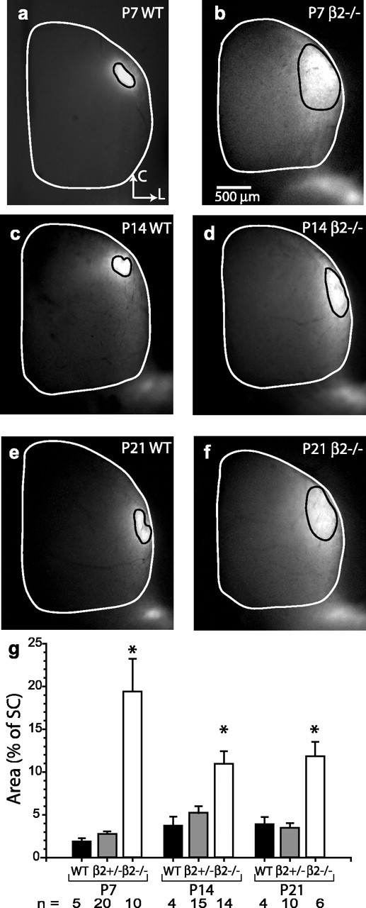
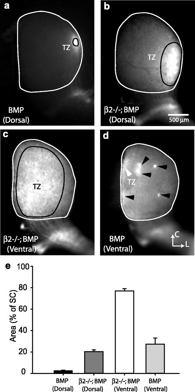
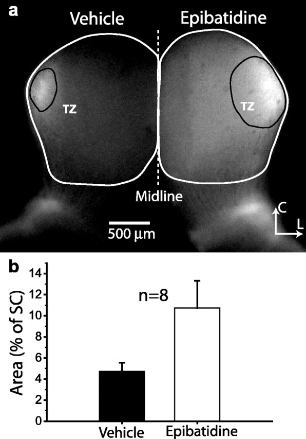
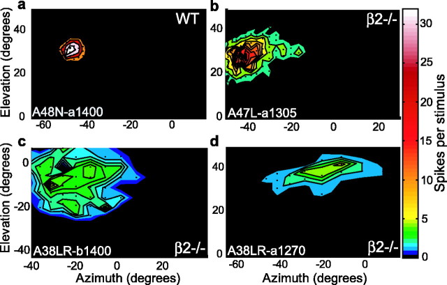
Although it is widely accepted that molecular mechanisms play an important role in the initial establishment of retinotopic maps, it has also long been argued that activity-dependent factors act in concert with molecular mechanisms to refine topographic maps. Evidence of a role for retinal activity in retinotopic map refinement in mammals is limited, and nothing is known about the effect of spontaneous retinal activity on the development of receptive fields in the superior colliculus. Using anatomical and physiological methods with two genetically manipulated mouse models and pharmacological interventions in wild-type mice, we show that spontaneous retinal waves instruct retinotopic map refinement in the superior colliculus of the mouse. Activity-dependent mechanisms may play a preferential role in the mapping of the nasal-temporal axis of the retina onto the colliculus, because refinement is particularly impaired along this axis in mutants without retinal waves. Interfering with both axon guidance cues and activity-dependent cues in the same animal has a dramatic cumulative effect. These experiments demonstrate how axon guidance cues and activity-dependent factors combine to instruct retinotopic map development.


Figures

Figure 1.
Figure 1.
β2-/- mice have enlarged retinocollicular projections. a, c, e, Examples of focal dye injections in the retina that result in focal spots of label (target zones; outlined in black) in the contralateral SC (outlined in white) of mice. Typical projections in P7, P14, and P21 WT SC. b, d, f, Typical projections in β2-/- mice at corresponding ages. g, Summary of quantified projection area for all ages and genotypes. WT (white bars) and heterozygous (β2+/-; gray bars) mice have similar projection areas at all time points.β2-/- (black bars) mice have larger projection ratios at all time points compared with WT or β2+/- mice. There is a partial rescue after the onset of glutamatergic retinal waves in the second week after birth but no additional rescue after the onset of visual experience at approximately P14 (t test; *p < 0.05). Error bars represent SEM.
Figure 2.
Figure 2.
Molecular and activity-dependent mechanisms combine to guide retinotopic map development in the SC. a, A focal retinal injection into the dorsal retina of a BMP transgenic mouse (P6-P7) results in a single normal confined target zone in lateral colliculus. b, In BMP transgenics that are also homozygous for the β2 mutation, dorsal retinal injections result in large target zones. c, A focal retinal injection into ventral retina of a BMP transgenic that is also homozygous for the β2 mutation results in a very large, completely unrefined pattern of projection to the SC. d, A ventral retinal injection into a BMP mutant results in a normal target zone in the medial colliculus (white arrowhead) as well as numerous ectopic spots (black arrowheads), typical of mutant mice with disturbed molecular cues responsible for establishing retinotopic maps in the colliculus. e, Quantification of the target zone area for the various genetic backgrounds and retinal injection sites. Dorsal injections into the BMP transgenic retina (black bar) have a completely normal target zone location and area, as is expected. Crossing the BMP mice onto a β2-/- background (β2-/-;BMP) causes an enlarged target zone, attributable to the absence of retinal waves (dark gray bar). Ventral injections into the retina of BMP mice on the β2-/- background (β2-/-;BMP) results in projections that nearly fill the SC (white bar). This demonstrates that focal ectopic spots, which normally result from ventral injections in BMP mice, are the result of the influence of retinal waves in the first week after birth [p < 0.0001 for difference between BMP (Dorsal); β2-/-;BMP (Dorsal), and β2-/-;BMP (Ventral)]. Error bars represent SEM. TZ, Target zone; C, caudal; L, lateral.
Figure 3.
Figure 3.
Epibatidine treatment to retina of WT mice mimics the phenotype of β2-/- whole-animal KO. a, Epibatidine was applied to one eye from P3-P7, with vehicle control treatment in the other eye. Focal dye injections at P7 result in larger projections to the SC contralateral to the epibatidine-treated eye than the SC contralateral to the vehicle-treated eye. Example of projections to the SC in a WT mouse from an eye treated with epibatidine (right) compared with projections to the SC in the same animal from an eye treated with vehicle solution (left). The labeled projection to the right SC, receiving input primarily from the epibatidine-treated eye, is significantly broader than the projection to the left SC, receiving input primarily from the vehicle-treated eye. b, Quantification from eight mice similarly treated in both eyes with either vehicle (black bar) or drug (white bar) reveals that epibatidine treatment results in larger projections from the retina to the SC (paired t test; p < 0.05). Error bars represent SEM. TZ, Target zone; C, caudal; L, lateral.
Figure 4.
Figure 4.
Enlarged in vivo RFs in the superior colliculus of β2-/- mutant mice. a, Typical multiunit receptive field in a WT mouse. The RF is compact, and response to stimulation of the RF center is large. b, Typical multiunit response in a β2-/- mouse. c, Example of a patchy receptive field of an isolated single neuron in a β2-/- mouse (Pearson's regression coefficient for 2D Gaussian of r2 = 0.32, indicating a very poor fit). d, Elongated RF in isolated β2-/- neuron. Mutant RFs are generally elongated along the nasal-temporal visual axis. Color scale bar on the right applies to all panels. Small black dots in each panel indicate spacing of visual stimuli.
Figure 5.
Figure 5.
Quantification of RF properties. a, Two-dimensional (elliptical) Gaussian fits, measured with the Pearson's regression coefficient, were significantly better in WT and β2+/- mice than in β2-/- mice for both multiunit and single-unit RFs (t test; p < 0.01). This suggests that β2-/- RFs are poorly organized and patchy compared with comparable WT and β2+/- mice. Graph shows the distribution of r2 values for each genotype along with means and SEs. b, Quantification of RF area (see Materials and Methods) reveals that multiunit mutant RFs (black bars) were much larger than corresponding WT (white bar) or β2+/- (dark gray bar) RFs (t test; **p < 0.01). Isolated single-unit RF areas in the combined population of WT and β2+/- mice were also much smaller than isolated single-unit β2-/- RF areas (t test; *p < 0.05). There was no difference between the WT and β2+/- multiunit data or between the multiunit and single-unit data within genotypes. c, Mutant RFs (black bars) typically had much lower peak response to visual stimuli than WT (white bar) and β2+/- (dark gray bar) mice. This is true for multiunits (white, dark gray, and black bars) as well as isolated single units (light gray and black bars; t test; **p < 0.01). d, The total stimulus response in WT, β2+/-, and β2-/- mice are similar. This suggests that the combination of the decrease in RF peak response and the increase in RF area results in no net change in input from the retina to the superior colliculus. Error bars represent SEM. MU, Multiunit; SU, single unit.
Figure 6.
Figure 6.
Retinocollicular projections are preferentially elongated along the NT axis of the retina in β2-/- mutant mice. a, Scatterplot of dorsoventral versus nasal-temporal RF projections in WT (open circles), β2+/- (gray circles), and β2-/- (black circles) mice. β2-/- RFs are larger and more elongated along the NT axis. Numbers include all well fit sites with multiunit response in addition to sites with only single-unit response. b, Paired comparisons of the NT and DV projections show a significant difference in the β2+/- and β2-/- mice (paired t test; WT, p = 0.06; β2+/-, *p = 0.02; β2-/-, **p ≪ 0.001). The projections along each axis are also significantly larger in the β2-/- compared with WT or β2+/- mice (t test; **p < 0.001). c, A similar measurement of the anatomical projections to the SC for P21 mice reveals a corresponding bias in the projections along the RC axis of the SC, which corresponds to the NT axis in visual space (paired t test; β2+/-, *p < 0.05; WT, *p < 0.01; β2-/-, **p < 0.001). Although the rostrocaudal projections of the mutant are significantly larger than the corresponding WT and β2+/- projections (t test; **p < 0.001), there is no difference between the mediolateral projections between genotypes. d, The ratio of the RC/ML anatomical projection in β2-/- mice is significantly higher than in WT (t test; **p < 0.01) or β2+/- (t test; **p < 0.001) mice. The ratio of the physiological response along the NT/DV axis is significantly different between WT and β2+/- mice (*p = 0.03) and highly significant between WT and β2-/- (**p ≪ 0.001) or between β2+/- and β2-/- (**p < 0.01) mice. This highlights the asymmetric effect of blocking retinal waves on mapping along the nasal-temporal axis of visual space. Error bars represent SEM.
Figure 7.
Figure 7.
Model for the combined effect of molecular and activity-dependent factors on retinotopic map development. Early in development (P1), retinal ganglion cells rely on axon guidance cues to target them selves to the colliculus. Illustrated are axons from dorsal (blue) and ventral (red) retina. In BMP transgenic mice (right column), these molecular cues are inappropriate for ventral axons; thus, their targeting in the colliculus is disrupted. During the first week after birth (between P1 and P8), retinal axons arborize preferentially along the rostrocaudal axis at a location determined by their nasal-temporal position in the retina. In BMP transgenic mice, the increased spread of ventral retinal axons along the mediolateral extent of the colliculus results in ventral arbors covering a large fraction of the colliculus. By P8, correlated retinal waves have refined the projection of retinal ganglion cell arbors to a retinotopically appropriate focal region in the colliculus. In the absence of retinal waves (left column, β2-/-), the projection pattern is left in an unrefined, immature state. In mutants with axon guidance defects but intact retinal waves (right column, β2+/+), activity-dependent Hebbian mechanisms mutually reinforce a small number of misprojecting (ventral) axons into refined focal ectopic spots. Dorsal retinal axons refine normally. In the absence of correlated activity in the retina (right column, β2-/-), dorsal (blue) axons in BMP transgenic mice target appropriately but do not refine into a focal spot. In the absence of both correlated activity and appropriate axon guidance cues, ventral (red) axons in the β2-/-;BMP mice make widespread projections that remain in an immature exuberant state. D, Dorsal; T, temporal; V, ventral; N, nasal; C, caudal; L, lateral; R, rostral; M, medial.

Similar articles

Cited by

References

    1. Baldi P, Heiligenberg W (1988) How sensory maps could enhance resolution through ordered arrangements of broadly tuned receivers. Biol Cybern 59: 313-318. - PubMed
    1. Bansal A, Singer JH, Hwang BJ, Xu W, Beaudet A, Feller MB (2000) Mice lacking specific nicotinic acetylcholine receptor subunits exhibit dramatically altered spontaneous activity patterns and reveal a limited role for retinal waves in forming ON and OFF circuits in the inner retina. J Neurosci 20: 7672-7681. - PMC - PubMed
    1. Carrasco MM, Razak KA, Pallas SL (2003) Effects of dark-rearing on retinocollicular map refinement. Soc Neurosci Abstr 29: 37.23.
    1. Chan SO, Guillery RW (1994) Changes in fiber order in the optic nerve and tract of rat embryos. J Comp Neurol 344: 20-32. - PubMed
    1. Chen C, Regehr WG (2000) Developmental remodeling of the retinogeniculate synapse. Neuron 28: 955-966. - PubMed

Publication types

MeSH terms

LinkOut - more resources