. 2005 Aug;3(8):e268.
doi: 10.1371/journal.pbio.0030268. Epub 2005 Jul 26.

De novo formation of left-right asymmetry by posterior tilt of nodal cilia

Affiliations

De novo formation of left-right asymmetry by posterior tilt of nodal cilia

Shigenori Nonaka et al. PLoS Biol. 2005 Aug.

Abstract

In the developing mouse embryo, leftward fluid flow on the ventral side of the node determines left-right (L-R) asymmetry. However, the mechanism by which the rotational movement of node cilia can generate a unidirectional flow remains hypothetical. Here we have addressed this question by motion and morphological analyses of the node cilia and by fluid dynamic model experiments. We found that the cilia stand, not perpendicular to the node surface, but tilted posteriorly. We further confirmed that such posterior tilt can produce leftward flow in model experiments. These results strongly suggest that L-R asymmetry is not the descendant of pre-existing L-R asymmetry within each cell but is generated de novo by combining three sources of spatial information: antero-posterior and dorso-ventral axes, and the chirality of ciliary movement.


Figures

Figure 1
Figure 1. Flow Generation Mechanism
Circular clockwise motion of a cilium can generate directional leftward flow if its axis is not perpendicular to the cell surface but tilted posteriorly. Due to distance from the cell surface, a cilium in the leftward phase (red arrow) drags surrounding water more efficiently than the rightward phase (blue arrow), resulting in leftward force in total (purple arrow). See text for details. The rotating cilium is drawn slightly bent due to viscous resistance, as seen in Video S1.
Figure 2
Figure 2. Trajectory of Node Cilia Movement
(A) Trace of node cilia in enhanced DIC images after background subtraction. Positions of root are indicated in black, and tip in blue, green, and orange. Most cilia have a pattern consistent with the projection of a tilted cone (blue and green, see text) whereas some cilia move in a D-shape (orange). A, P, L, and R refer to anterior, posterior, left, and right sides of the node, respectively. The direction of cilia rotation was clockwise (arrows). (B) Relationship between essentially rotatory movement of cilia and their projected images at various tilt angles.
Figure 3
Figure 3. Node Cilia Are Posteriorly Tilted and Positioned
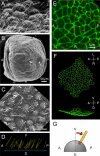
(A) Scanning electron micrograph of the wild-type node. Note that cilia emanate from the posterior part of the cells. The view angle is about 30° with respect to the horizontal line. (B, C) Scanning electron micrograph of the iv/iv node. (C) is a high-magnification picture of the region in (B) indicated by an arrow. (D) Deduced tilt of iv/iv node cilia after stereography from multiple-tilt scanning electron micrograph images. Yellow lines indicate cilia, red dots their root positions, and a blue square a plane best-fit to the node surface. When we calculated the tilt of the cilia, we separated the tilt into A-P (anterior–posterior) and L-R components. The average tilt was 26.6° in A-P axis (toward the posterior) and 0.06° in L-R axis (towards the right). (E) Immunofluorecence image of node cells shown as projection of 3D confocal data stack. Basal bodies and cell boundaries are shown by immunofluorescence against γ-tubulin (red) and ZO-1 (green), respectively. (F) 3D reconstruction of (E) viewed from ventral side (top) and right side (bottom), showing posterior bias of basal body positions. White lines divide the cells into the anterior and the posterior halves. Basal bodies located in the anterior and the posterior are shown in yellow and red, respectively. (G) Speculative interpretation of posterior bias of basal bodies in orientation and position of the node cilia. Because the node cells are somewhat rounded, if basal bodies were located at the posterior part of these cells, it would result in posteriorly tilted cilia even though the basal bodies remain perpendicular to the plasma membrane.
Figure 4
Figure 4. Experimental Fluid Dynamics Model of the Posterior Tilt Mechanism
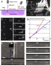
(A) Schematic representation of the model. Wires driven by stepper motors stir silicone fluid of 30,000 centipoise. Note that the fluid surface is in contact with the acrylic board. Resulting flow is visualized as movement of glitter particles inserted through holes on the board. Wires are connected to motor axles by elastomer connectors in order to smoothen the stepwise motion of the motors. The path of the wires is completely specified by the two angles θ and ψ, as shown in the diagram. (B) Photograph of the model. Corresponding directions are indicated. (C) Leftward flow. Flow generated by five rotating wires (visible as white lines in photo) drives glitter towards the left at the depth of 3 mm from the surface. Trajectory of the glitter becomes curved far from the wires (t = 3′) because the glitter has reached the chamber's wall. Scale = 1 cm. (D) Flow velocity (leftward component) as a function of tilt and bend angles. The flow velocity shown here represents the flow speed at the depth of 3 mm. The flow is most efficient when the path of the wire is tangential to the board surface during part of its rotation (i.e., ψ + θ = 90°). (E) Leftward flow near the surface. Glitter was injected on the right side at the surface of the fluid (no more than 1-mm depth). Only leftward flow was observed. Scale = 1 cm.

References

    1. Hamada H, Meno C, Watanabe D, Saijoh Y. Establishment of vertebrate left-right asymmetry. Nat Rev Genet. 2002;3:103–113. - PubMed
    1. Nonaka S, Tanaka Y, Okada Y, Takeda S, Harada A. Randomization of left-right asymmetry due to loss of nodal cilia generating leftward flow of extraembryonic fluid in mice lacking KIF3B motor protein. Cell. 1998;95:829–837. - PubMed
    1. Nonaka S, Shiratori H, Saijoh Y, Hamada H. Determination of left-right patterning of the mouse embryo by artificial nodal flow. Nature. 2002;418:96–99. - PubMed
    1. Tabin CJ, Vogan KJ. A two-cilia model for vertebrate left-right axis specification. Genes Dev. 2003;17:1–6. - PubMed
    1. McGrath J, Somlo S, Makova S, Tian X, Brueckner M. Two populations of node monocilia initiate left-right asymmetry in the mouse. Cell. 2003;114:61–73. - PubMed

Publication types