. 2005 Jun;7(6):623-9.
doi: 10.1593/neo.04781.

Brain tumor tropism of transplanted human neural stem cells is induced by vascular endothelial growth factor

Affiliations

Brain tumor tropism of transplanted human neural stem cells is induced by vascular endothelial growth factor

Nils Ole Schmidt et al. Neoplasia. 2005 Jun.

Abstract

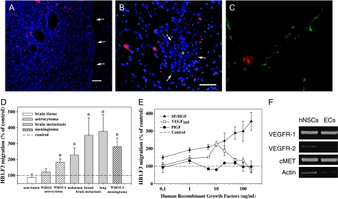
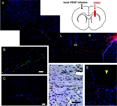
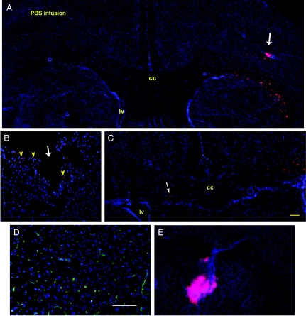
The transplantation of neural stem cells (NSCs) offers a new potential therapeutic approach as a cell-based delivery system for gene therapy in brain tumors. This is based on the unique capacity of NSCs to migrate throughout the brain and to target invading tumor cells. However, the signals controlling the targeted migration of transplanted NSCs are poorly defined. We analyzed the in vitro and in vivo effects of angiogenic growth factors and protein extracts from surgical specimens of brain tumor patients on NSC migration. Here, we demonstrate that vascular endothelial growth factor (VEGF) is able to induce a long-range attraction of transplanted human NSCs from distant sites in the adult brain. Our results indicate that tumor-upregulated VEGF and angiogenic-activated microvasculature are relevant guidance signals for NSC tropism toward brain tumors.


Figures

Figure 1
Figure 1
Brain tumor tropism of the human neural stem cell line, HB1.F3. (A) Distribution of migrated NSCs (red) within the tumor mass of a U87 human glioblastoma xenograft (arrows = tumor border; blue = cell nuclei; bar = 150 µm). (B) Invaded tumor cells distant from the main tumor mass of a U251 human glioblastoma xenograft coopting a blood vessel were surrounded by human NSCs (* = blood vessel; bar = 100 µm). (C) Colocalization of an NSC (red) with a CD31-positive tumor blood vessel (green) within a U87 human glioblastoma. (D) Effects of protein extracts from different human brain tumor specimens normalized to 400 µg/ml protein on human NSC migration in the modified Boyden chamber assay. Values shown are mean ± SE and are expressed as percentage of the unstimulated basal migratory rate (control = 100%) (*P < .01, Mann-Whitney U test). (E) Recombinant human VEGF165, PlGF, and SF/HGF were tested in a modified 96-well Boyden chamber assay. SF/HGF induced a higher chemotactic response than VEGF. No significant effects were seen for PlGF. (F) Human NSCs expressed the receptor mRNA of VEGFR-2 and VEGFR-1 for VEGF and PlGF, and cMET for SF/HGF. Endothelial cells (PAE/KDR) known to express all three different receptors were used as a positive control.
Figure 2
Figure 2
Representative sections of VEGF-induced migration of human NSCs HB1.F3 in the brain of adult nude mice (see diagram). (A) Long-range attraction of NSCs (red) from the right hemisphere (R) across the corpus callosum (cc) in response to a local microinfusion of VEGF (0.25 ng/hr) in the left hemisphere (L) (day 9; blue = cell nuclei). (B) Colocalization of NSCs with dilated CD31-positive (green) blood vessels near the VEGF infusion cannula (bar = 50 µm). (C) Already after 5 days, transhemispheric migrated NSCs pool in the area of VEGF infusion (bar = 100 µm). (D) Migrating NSCs (brown) in the corpus callosum as demonstrated by immunohistochemistry using a human-specific antibody (bar = 25 µm). (E) Local microinfusion of SF/HGF (0.25 ng/hr) was not able to induce a long-range attraction. Only directly at the injury site of the cannula (arrowhead) were some NSCs observed.
Figure 3
Figure 3
(A–C) Representative coronal brain sections of different nude mice receiving a constant intraparenchymal PBS infusion in the left hemisphere. (A) No extensive migration toward the PBS-infused left hemisphere was observed 9 days after injection of Dil-labeled human NSCs HB1.F3 (red) in the right forebrain (arrow). (B) However, some NSCs (arrowheads) were found at the lesion site of the brain cannula (arrow) and (C) occasionally single NSCs (arrow) migrating to the opposite infused hemisphere could be detected. This indicates that already the injury induced by the infusion cannula triggers mechanisms causing minor NSC migration (bar = 50 µm). (D) SF/HGF, although known to display angiogenic properties, did not cause a stimulation of angiogenesis near the cannula when infused at the dosage of 0.25 ng/hr (CD31 immunoreactivity = green) (bar = 100 µm). (E) In control experiments, Dil-labeled endothelial cells (red) stayed at the site of injection and did not migrate toward the VEGF infusion site.

References

    1. Kleihues P, Cavenee WK. Pathology and Genetics of Tumors of the Central Nervous System International Agency for Research on Cancer Press, Lyon. 2000.
    1. Aboody KS, Brown A, Rainov NG, Bower KA, Liu S, Yang W, Small JE, Herrlinger U, Ourednik V, Black PM, et al. From the cover: neural stem cells display extensive tropism for pathology in adult brain: evidence from intracranial gliomas. Proc Natl Acad Sci USA. 2000;97:12846–12851. - PMC - PubMed
    1. Benedetti S, Pirola B, Pollo B, Magrassi L, Bruzzone MG, Rigamonti D, Galli R, Selleri S, Di Meco F, De Fraja C, et al. Gene therapy of experimental brain tumors using neural progenitor cells. Nat Med. 2000;6:447–450. - PubMed
    1. Ehtesham M, Kabos P, Kabosova A, Neuman T, Black KL, Yu JS. The use of interleukin 12-secreting neural stem cells for the treatment of intracranial glioma. Cancer Res. 2002;62:5657–5663. - PubMed
    1. Staflin K, Honeth G, Kalliomaki S, Kjellman C, Edvardsen K, Lindvall M. Neural progenitor cell lines inhibit rat tumor growth in vivo. Cancer Res. 2004;64:5347–5354. - PubMed

Publication types

MeSH terms

Substances

LinkOut - more resources