. 2005 Aug 16;102(33):11581-8.
doi: 10.1073/pnas.0504592102. Epub 2005 Jul 21.

Modulation of disease, T cell responses, and measles virus clearance in monkeys vaccinated with H-encoding alphavirus replicon particles

Affiliations

Modulation of disease, T cell responses, and measles virus clearance in monkeys vaccinated with H-encoding alphavirus replicon particles

Chien-Hsiung Pan et al. Proc Natl Acad Sci U S A. .

Abstract

Measles remains a major worldwide problem partly because of difficulties with vaccination of young infants. New vaccine strategies need to be safe and to provide sustained protective immunity. We have developed Sindbis virus replicon particles that express the measles virus (MV) hemagglutinin (SIN-H) or fusion (SIN-F) proteins. In mice, SIN-H induced high-titered, dose-dependent, MV-neutralizing antibody after a single vaccination. SIN-F, or SIN-H and SIN-F combined, induced somewhat lower responses. To assess protective efficacy, juvenile macaques were vaccinated with a single dose of 10(6) or 10(8) SIN-H particles and infant macaques with two doses of 10(8) particles. A dose of 10(8) particles induced sustained levels of high-titered, MV-neutralizing antibody and IFN-gamma-producing memory T cells, and most monkeys were protected from rash when challenged with wild-type MV 18 months later. After challenge, there was a biphasic appearance of H- and F-specific IFN-gamma-secreting CD4+ and CD8+ T cells in vaccinated monkeys, with peaks approximately 1 and 3-4 months after challenge. Viremia was cleared within 14 days, but MV RNA was detectable for 4-5 months. These studies suggest that complete clearance of MV after infection is a prolonged, phased, and complex process influenced by prior vaccination.


Figures

Fig. 1.
Fig. 1.
MV-neutralizing antibody induced in BALB/c mice by SINV replicon vaccines. Shown are responses induced by SIN-H, SIN-F, and SIN-H plus SIN-F particles 4 weeks after i.d. or i.m. inoculation with a single dose of 104,105, or 106 particles. Data are presented as the GMT of two to three individual mice. *, P < 0.05; ***, P < 0.005; and ****, P < 0.001, compared with the next lower dose. PRNT, 50% plaque reduction neutralization titer.
Fig. 2.
Fig. 2.
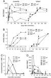
Immune responses induced by vaccination of juvenile and infant monkeys with SIN-H particles. (A) MV-neutralizing antibody measured by 50% plaque reduction in Vero cells (PRNT). Dashed horizontal line indicates the generally accepted protective level. (B) Avidity of MV-specific EIA antibody measured by resistance to dissociation with increasing concentrations of sodium thiocyanate. (C) H-specific cellular immune responses measured by lymphocyte proliferation to H peptides. (D) SINV-specific neutralizing antibody measured by 50% plaque reduction in BHK cells. Arrows indicate the time of boost for infant macaques 23P and 37P.
Fig. 3.
Fig. 3.
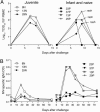
Protection from challenge with wild-type MV as determined by rash, level of viremia, and induction of IgM antibody. (A) Viremia as measured by cocultivation of PBMC with B95-8 cells. Monkeys that developed a rash are indicated. (B) MV-specific IgM as measured by EIA expressed as optical density (OD) for plasma diluted 1:200.
Fig. 4.
Fig. 4.
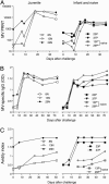
Characteristics of antibody responses after challenge as measured by virus neutralization and EIA. Plasma samples were collected at various times after challenge and measured for MV neutralization by plaque reduction on Vero cells (A), the absorbance (OD) of MV-specific IgG at 1:400 dilution of plasma by EIA (B), and the avidity of the IgG EIA antibody measured by resistance to dissociation with increasing concentrations of sodium thiocyanate (C). For avidity, points represent the mean and standard error for multiple dilutions of plasma.
Fig. 5.
Fig. 5.
IFN-γ production by MV-specific T cells in peripheral blood after challenge. PBMCs were assayed at various times after challenge for IFN-γ ELISPOT formation in response to stimulation with pooled MV H peptides (A) or MV F peptides (B). PBMCs collected 35 and 125 days after challenge were depleted of CD4 T cells, and the responses of depleted and nondepleted cells to pools of H (C) and F (D) peptides were compared.

Similar articles

Cited by

References

    1. Centers for Disease Control (2003) Morbid. Mortal. Wkly. Rep. 52, 471–475. - PubMed
    1. Muscat, M., Glismann, S. & Bang, H. (2003) Euro. Surveill. 8, 123–129. - PubMed
    1. Moss, W. J., Monze, M., Ryon, J. J., Quinn, T. C., Griffin, D. E. & Cutts, F. (2002) Clin. Infect. Dis. 35, 189–196. - PubMed
    1. Cutts, F. T., Henao-Restrepo, A. & Olive, J. M. (1999) Vaccine 17, Suppl. 3, S47–S52. - PubMed
    1. Centers for Disease Control (2000) Morbid. Mortal. Wkly. Rep. 49, 1116–1118. - PubMed

Publication types

MeSH terms