. 2005 Aug 5;97(3):219-26.
doi: 10.1161/01.RES.0000177862.85474.63. Epub 2005 Jul 21.

Alk3/Bmpr1a receptor is required for development of the atrioventricular canal into valves and annulus fibrosus

Affiliations

Alk3/Bmpr1a receptor is required for development of the atrioventricular canal into valves and annulus fibrosus

Vinciane Gaussin et al. Circ Res. .

Abstract

Endocardial cushions are precursors of mature atrioventricular (AV) valves. Their formation is induced by signaling molecules originating from the AV myocardium, including bone morphogenetic proteins (BMPs). Here, we hypothesized that BMP signaling plays an important role in the AV myocardium during the maturation of AV valves from the cushions. To test our hypothesis, we used a unique Cre/lox system to target the deletion of a floxed Alk3 allele, the type IA receptor for BMPs, to cardiac myocytes of the AV canal (AVC). Lineage analysis indicated that cardiac myocytes of the AVC contributed to the tricuspid mural and posterior leaflets, the mitral septal leaflet, and the atrial border of the annulus fibrosus. When Alk3 was deleted in these cells, defects were seen in the same leaflets, ie, the tricuspid mural leaflet and mitral septal leaflet were longer, the tricuspid posterior leaflet was displaced and adherent to the ventricular wall, and the annulus fibrosus was disrupted resulting in ventricular preexcitation. The defects seen in mice with AVC-targeted deletion of Alk3 provide strong support for a role of Alk3 in human congenital heart diseases, such as Ebstein's anomaly. In conclusion, our mouse model demonstrated critical roles for Alk3 signaling in the AV myocardium during the development of AV valves and the annulus fibrosus.


Figures
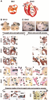
Figure 1
Figure 1
Fate of the AV myocardial lineage. A, AV valve development (see text). Red, myocardium; Orange, endocardium; Yellow, mesenchyme. B and C, Whole-mount LacZ staining of hearts from E11.5 and E14.5 cGATA6-Cre/R26R mice. Arrow, posterior right AVC. Star, left AVC. D through EE, After whole-mount LacZ staining of hearts from cGATA6-Cre/R26R mice, sections were immunostained with the MF20 antibody to identify cardiac myocytes (brown) (D through N; AA through CC) or counterstained with the Van Gieson stain to identify the AV valves and annulus fibrosus (pink) (O through Z; DD and EE). Arrowheads, LacZ-positive cells. L, cushion-derived mesenchyme (yellow arrow) and epicardially-derived sulcus tissue (star) fuse to form the annulus fibrosus. Scale bar, 500 μm (O, R, U, X, and DD), 200 μm (G, K, and EE), 100 μm (D and AA), or 50 μm (remaining sections). A indicates atrium; AVC, AV canal; ECC, endocardial cushion; iECC, inferior ECC; IVS, interventricular septum; LV, left ventricle; mml, mitral mural leaflet; msl, mitral septal leaflet; OT, outflow tract; RA, right atrium; RV, right ventricle; sECC, superior ECC; tml, tricuspid mural leaflet; tpl, tricuspid posterior leaflet; tsl, tricuspid septal leaflet; V, ventricle.
Figure 2
Figure 2
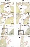
Histological analysis of cGATA6-Cre/Alk3 mice. Coronal (A through D) and sagital (E through H) sections through the heart of 3-month-old cGATA6-Cre/Alk3 mice (B, D, F, and H) and control littermates (Alk3null/flox) (A, C, E, and G) were stained with Picric Acid Sirius Red and revealed morphological anomalies in the mitral septal leaflet (B) and tricuspid mural leaflet (D). There was a downward displacement of the posterior annulus fibrosus (compare position of arrowheads in E and F, which indicate the annulus fibrosus). Arrow and dotted line in F indicate where the annulus fibrosus was expected. The tricuspid posterior leaflet was adherent to the ventricular wall and contained nonfibrous tissue (star in H). Scale bar, 500 μm (A through F) or 200 μm (G and H). IVS, interventricular septum.
Figure 3
Figure 3
Premature disappearance of the myocardial layer in the tricuspid mural leaflet in the absence of Alk3. Sections through the heart of E14.5 (A and B) and E15.5 embryos (C and D) were stained with MF20 antibody (brown) and counterstained with hematoxylin (blue). A and C, Coronal section through the heart of a control embryo (Alk3null/flox) showing a homogenous layer of cardiac myocyte (arrow) in the tricuspid mural leaflet. B and D, In cGATA6-Cre/Alk3 embryos, this layer is present at E14.5 (arrow; B) but missing at E15.5 (arrowhead; D). Star, papillary muscle (C and D). Scale bar, 100 μm.
Figure 4
Figure 4
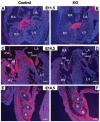
Alk3 is required for periostin expression in the developing AV valves. In situ hybridization for periostin in E11.5 (A and B) and E14.5 (C and F) embryos. Robust expression of periostin was seen in the mesenchymal cells of the endocardial cushions in E11.5 (A) and E14.5 (C) control embryos (Alk3null/flox). Star, myocardial layer in the tricuspid mural leaflet (C). In cGATA6-Cre/Alk3 embryos, similar periostin expression was detected in the endocardial cushion at E11.5 (B) but was dramatically decreased in both AV valves at E14.5 (D), whereas expression outside the heart, ie, within the periosteum of the ribs, was unaffected (E and F). The positive signal (pink) overlays Hoechst nuclear staining (blue). Scale bar, 100 μm. R, indicates rib.
Figure 5
Figure 5
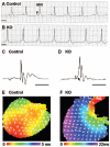
ECG analysis and optical mapping of hearts from cGATA6-Cre/Alk3 mice. Representative surface ECG (lead I) from 6-month-old cGATA6-Cre/Alk3 mice (B) showing short PR interval when compared with control littermate (Alk3null/flox) (A). Scale bar, 50 ms. C and D, Volume-conducted electrocardiograms were recorded in isolated Langendorff perfused hearts and showed the presence of a delta wave (arrow) in cGATA6-Cre/Alk3 mice (D) when compared with control littermate (Alk3null/flox) (C). Scale bar, 50 ms. E and F, High-resolution optical mapping with the voltage sensitive dye Di-4-ANEPPS was recorded in isolated Langendorff perfused hearts from the posterior side while the hearts were beating spontaneously in sinus rhythm. Control hearts (Alk3null/flox) showed a mature apex-to-base ventricular activation pattern (E), whereas in hearts with abnormal surface ECG, a base-to-apex activation pattern was observed, with evidence of a posterior paraseptal bypass tract (F).
Figure 6
Figure 6
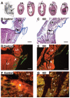
Histological analysis of the bypass tract in cGATA6-Cre/Alk3 mice. A, Serial sagital sections were obtained through the heart of 3-month-old control heart and stained with Masson’s trichrome. B, Higher magnification of the posterior paraseptal region outlined in A showing insulation of the atrium from the ventricle by the annulus fibrosus (arrow). C, Same area as in B, but in a heart from a cGATA6-Cre/Alk3 mouse showing disruption of the annulus fibrosus and myocytic connection between the atrium and the ventricle (arrow). Immunofluorescence on adjacent sections for periostin (green; D and E) or Cx43 (green; F and G) and α-cardiac actinin (red). Periostin is localized in the annulus fibrosus in a control heart (arrow; D) but is not detectable in that area in the heart with the bypass tract (arrow; E). Cx43 is present in the cardiac myocytes of the bypass tract (arrowheads; G), whereas it is not detected in the annulus fibrosus (star) in a control heart (F). F and G, Adjacent sections to the boxed area in B and C, respectively. Scale bar, 100 μm (B through E).
Figure 7
Figure 7
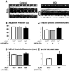
Echocardiographic analysis of cGATA6-Cre/Alk3 mice. A, Representative echocardiographic recordings in a 3-month-old control (Alk3null/flox) (left) and a cGATA6-Cre/Alk3 mouse (right). B through D, Three-month-old cGATA6-Cre/Alk3 mice (KO) had a significant decrease in LV ejection fraction (B) and a significant increase in LV end systolic dimension (C) and LV end diastolic dimension (D) when compared with control littermates (Alk3null/flox). E, LV dysfunction was accompanied by increased expression of ANF in the myocardium as measured by quantitative RT-PCR. *P<0.05.

References

    1. Hoffman J. Incidence of congenital heart disease: II. Prenatal incidence. Pediatr Cardiol. 1995;16:155–165. - PubMed
    1. Eisenberg LM, Markwald RR. Molecular regulation of atrioventricular valvuloseptal morphogenesis. Circ Res. 1995;77:1–6. - PubMed
    1. Markwald R, Eisenberg C, Eisenberg L, Trusk T, Sugi Y. Epithelial-mesenchymal transformations in early avian heart development. Acta Anat (Basel) 1996;156:173–186. - PubMed
    1. Schroeder JA, Jackson LF, Lee DC, Camenisch TD. Form and function of developing heart valves: coordination by extracellular matrix and growth factor signaling. J Mol Med. 2003;81:392–403. - PubMed
    1. Barnett JV, Desgrosellier JS. Early events in valvulogenesis: a signaling perspective. Birth Defects Res C Embryo Today. 2003;69:58–72. - PubMed

Publication types