Comparative Study
. 2005 Aug 17;24(16):2862-72.
doi: 10.1038/sj.emboj.7600752. Epub 2005 Jul 28.

Nuclear export and cytoplasmic processing of precursors to the 40S ribosomal subunits in mammalian cells

Affiliations
Comparative Study

Nuclear export and cytoplasmic processing of precursors to the 40S ribosomal subunits in mammalian cells

Jacques Rouquette et al. EMBO J. .

Abstract

It is generally assumed that, in mammalian cells, preribosomal RNAs are entirely processed before nuclear exit. Here, we show that pre-40S particles exported to the cytoplasm in HeLa cells contain 18S rRNA extended at the 3' end with 20-30 nucleotides of the internal transcribed spacer 1. Maturation of this pre-18S rRNA (which we named 18S-E) involves a cytoplasmic protein, the human homolog of the yeast kinase Rio2p, and appears to be required for the translation competence of the 40S subunit. By tracking the nuclear exit of this precursor, we have identified the ribosomal protein Rps15 as a determinant of preribosomal nuclear export in human cells. Interestingly, inhibition of exportin Crm1/Xpo1 with leptomycin B strongly alters processing of the 5'-external transcribed spacer, upstream of nuclear export, and reveals a new cleavage site in this transcribed spacer. Completion of the maturation of the 18S rRNA in the cytoplasm, a feature thought to be unique to yeast, may prevent pre-40S particles from initiating translation with pre-mRNAs in eukaryotic cells. It also allows new strategies for the study of preribosomal transport in mammalian cells.


Figures

Figure 1
Figure 1
Pre-rRNA processing in HeLa cells. Schematic and nomenclature according to Hadjiolova et al (1993). Two alternative pathways are presented.
Figure 2
Figure 2
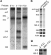
A new late precursor to the 18S rRNA in HeLa cells, the 18S-E pre-rRNA. (A) Northern blot analysis of total RNA extracts from HeLa cells. Northern blots probed with oligonucleotides complementary to the ETS1, the 5′ of the ITS1 (5′-ITS1) and the ITS2. The 5′-ITS1 probe hybridizes with a rapidly migrating precursor of the 18S rRNA, which we called 18S-E. (B) Synthesis of the 18S-E pre-rRNA is sensitive to treatment with actinomycin D. Analysis by Northern blot (probes: 5′-ITS1 and actin) and ethidium bromide staining (18S and 28S RNAs) of total RNA extracts from HeLa cells treated for 1 h with 40 ng/ml actinomycin D. (C) Northern blot analysis of total RNA extracts from HeLa cells probed with the 5′-ITS1 and 5687-ITS1 oligonucleotides.
Figure 3
Figure 3
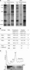
The 18S-E pre-rRNA is mostly located in the cytoplasm and does not participate in translation. (A) Northern blot analysis of total, cytoplasmic and nuclear RNA extracts from human HeLa cells and murine L929 cells shows localization of the 18S-E pre-rRNA to the cytoplasm. Hybridization was performed with probes complementary to the 5′-most nucleotides of the ITS1 and specific to each species. Only 1/10th of the total and cytoplasmic extracts was loaded on the gel. (B) Intracellular distribution of the 18S-E, 18S and 28S RNAs under various conditions was quantified on Northern blots by phosphorimaging. n: number of experiments. (C) Ribosome/polysome separation on sucrose gradient and Northern blot analysis of the fractions. HeLa cell cytoplasmic extracts were centrifuged on a 10–50% sucrose gradient. The 18S and 28S rRNAs were detected by ethidium bromide staining and the 18S-E species was revealed by Northern blotting with the 5′-ITS1 probe. The 18S-E RNA is found with the free 40S subunits.
Figure 4
Figure 4
Localization of hRio2 in HeLa cells. (A) Visualization of HeLa cells expressing Rio2-EGFP and EGFP by laser-scanning confocal microscopy. (B) Western blot of the nucleus and cytoplasm fractions probed with anti-GFP antibodies shows that hRio2-GFP is a cytoplasmic protein. As a control, the histone deacetylase HDAC2 is detected in the nuclear fraction. (C) hRio2-GFP is actively exported from the nucleus through a mechanism depending on Crm1, but not on preribosome synthesis. At 48 h after transfection with the hRio2-EGFP expression vector, cells were treated with 10 nM LMB for 2 h, or with 0.04 μg/ml actinomycin D for 1 h. (D) A fraction of hRio2-GFP cosediments with (pre-)40S particles on a sucrose gradient. Lysates from cells expressing hRio2-EGFP or EGFP were fractionated by ultracentrifugation on sucrose gradient as in Figure 3B. Fractions were analyzed by SDS–PAGE and Western blot with antibodies against GFP and against S19, a protein of the 40S subunit which is used as a marker of the free (pre-)40S subunits, the 80S monosomes and the polysomes. Note that expression of EGFP was much higher than that of hRio2-EGFP.
Figure 5
Figure 5
Analysis of pre-rRNA processing in HeLa cells after knockdown of hRio2 expression with siRNAs. (A) Northern blot analysis with the 5′-ITS1 probe of total RNAs shows accumulation of the 18S-E pre-rRNA in HeLa cells treated with hRio2 siRNAs. A scrambled siRNA with no defined target was used as a control. Equal amounts of RNA were loaded in each lane. The intensity profiles were determined by phophorimaging and normalized to the amount of 28S rRNA. The levels of 18S and 28S rRNAs were measured by reprobing the blot with complementary oligonucleotides (not shown). The values of the ratios were arbitrarily set to 1.0 in control cells. (B) Polysome analysis of HeLa cells treated with scrambled or hRio2 siRNAs and detection by Northern blot of the 18S-E pre-rRNA. Depletion of hRio2 leads to a strong imbalance between the free 60S and 40S subunits, indicating a defect in 40S subunit production. The 18S and 28S rRNAs were visualized with ethidium bromide. (C) FISH with the 5′-ITS1 probe conjugated to Cy3 shows accumulation of 18S-E pre-rRNA in the cytoplasm. In parallel, the U2 snRNA was detected with a Cy5-labeled probe. Quantification of the fluorescence was performed as described in Materials and methods. For the 5′-ITS1 probe, the results of two separate experiments are shown (***P<0.001, Student's t-test). (D) Northern blot analysis of subcellular nuclear and cytoplasmic fractions of HeLa cells with the 5′-ITS1 probe reveals accumulation of the 18S-E pre-rRNA in the cytoplasm upon treatment with hRio2 siRNA for 72 h. Only 1/10th of the cytoplasmic fraction was loaded on the gel. The levels of 18S and 28S were determined on the same blot with specific probes. The subcellular distribution of the 18S-E, 18S and 28S RNAs was quantified by phosphorimaging. N: nucleus; C: cytoplasm.
Figure 6
Figure 6
Characterization of nuclear export factors for the pre-40S particles. (A) Rps15 siRNAs specifically downregulate the level of Rps15 mRNA and efficiently block the production of the 40S subunit. RT–PCR analysis showed that treatment with Rps15 siRNAs for 48 h strongly affected the level of the Rps15 mRNA, but not that of the Rps19 mRNA (inset). As seen by polysome analysis on sucrose gradients, knockdown of Rps15 expression resulted in the disappearance of the free 40S subunit pool and in a strong increase in the level of free 60S subunits (compare with the control profiles in Figures 3 and 5). The 18S-E pre-rRNA was detected at very low levels in the fractions by Northern blot. The 18S and 28S rRNAs were visualized by ethidium bromide staining. (B) Northern blot analysis of total RNAs from HeLa cells treated with siRNAs directed against hRps15 or with LMB. Hybridization performed with the 5′-ITS1 probe. The intensity profiles were established as in Figure 5A. The levels of 18S and 28S rRNAs were measured by reprobing the blot with complementary oligonucleotides (not shown). The values of the ratios were arbitrarily set to 1.0 in control cells. (C) Detection by Northern blot of the 18S-E pre-rRNA in subcellular fractions of HeLa cells treated with siRNAs against Rps15 or with LMB. Distribution of the 18S-E pre-rRNA was quantified by phosphorimaging. N: nucleus; C: cytoplasm. (D) FISH with the 5′-ITS1 probe on HeLa cells treated with siRNAs against Rps15 or with LMB.
Figure 7
Figure 7
Characterization of the pre-rRNAs accumulating in LMB-treated cells. (A) Metabolic labeling of RNAs in HeLa cells with [32P]orthophosphate. Time after addition of [32P]orthophosphate to the medium is indicated (see Materials and methods). Total RNAs were extracted, separated by electrophoresis, and revealed by autoradiography. (B) Mapping of the extremities of the 26S RNA with probes complementary to the 5′-ETS (ETS1) and to the ITS1. Comparison of total RNAs from untreated and LMB-treated cells facilitates visualization of the 26S RNA. (C) Determination of the 5′-end of the 26S RNA by primer extension. Reverse transcription was performed with oligonucleotide ETS1-1722 on nuclear RNAs from LMB-treated cells. (D) Proposed boundaries for the 26S and 43S pre-rRNAs.
Figure 8
Figure 8
Transport and maturation of the 18S-E pre-rRNA. Crm1 may be implicated in nuclear export of the pre-40S subunits, but is also required for pre-18S rRNA maturation. Nuclear export of the pre-40S subunits requires Rps15, whereas hRio2 is necessary for conversion to mature 40S subunits.

References

    1. Beltrame M, Tollervey D (1992) Identification and functional analysis of two U3 binding sites on yeast pre-ribosomal RNA. EMBO J 11: 1531–1542 - PMC - PubMed
    1. Bohnsack MT, Regener K, Schwappach B, Saffrich R, Paraskeva E, Hartmann E, Gorlich D (2002) Exp5 exports eEF1A via tRNA from nuclei and synergizes with other transport pathways to confine translation to the cytoplasm. EMBO J 21: 6205–6215 - PMC - PubMed
    1. Borovjagin AV, Gerbi SA (2001) Xenopus U3 snoRNA GAC-Box A′ and Box A sequences play distinct functional roles in rRNA processing. Mol Cell Biol 21: 6210–6221 - PMC - PubMed
    1. Boulon S, Verheggen C, Jady BE, Girard C, Pescia C, Paul C, Ospina JK, Kiss T, Matera AG, Bordonne R, Bertrand E (2004) PHAX and CRM1 are required sequentially to transport U3 snoRNA to nucleoli. Mol Cell 16: 777–787 - PubMed
    1. Eichler DC, Craig N (1994) Processing of eukaryotic ribosomal RNA. Prog Nucleic Acid Res Mol Biol 49: 197–239 - PubMed

Publication types

MeSH terms