. 2006 Mar;27(3):213-29.
doi: 10.1002/hbm.20178.

Spatiotemporal mapping of cortical activity accompanying voluntary movements using an event-related beamforming approach

Affiliations

Spatiotemporal mapping of cortical activity accompanying voluntary movements using an event-related beamforming approach

Douglas Cheyne et al. Hum Brain Mapp. 2006 Mar.

Abstract

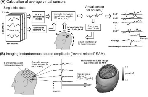
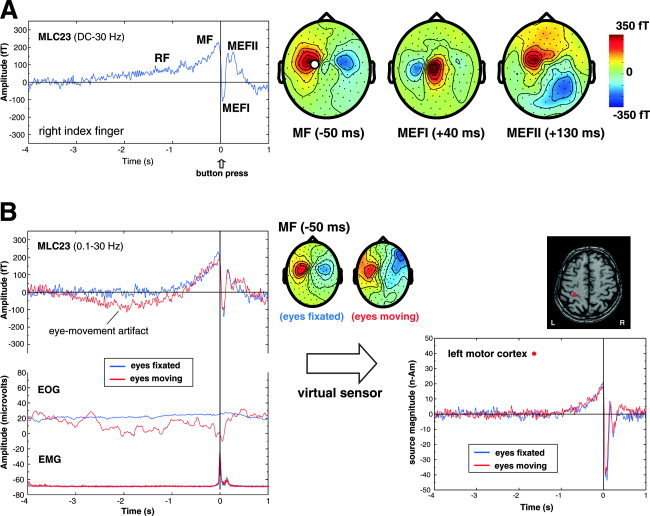
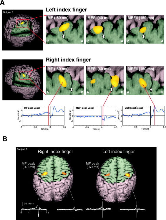
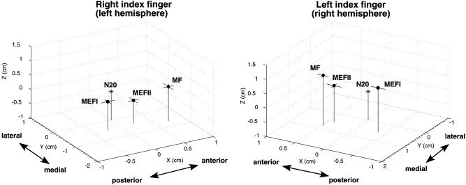
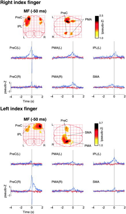
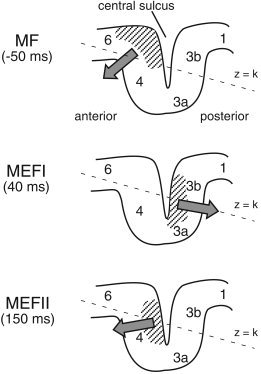
We describe a novel spatial filtering approach to the localization of cortical activity accompanying voluntary movements. The synthetic aperture magnetometry (SAM) minimum-variance beamformer algorithm was used to compute spatial filters three-dimensionally over the entire brain from single trial neuromagnetic recordings of subjects performing self-paced index finger movements. Images of instantaneous source power ("event-related SAM") computed at selected latencies revealed activation of multiple cortical motor areas prior to and following left and right index finger movements in individual subjects, even in the presence of low-frequency noise (e.g., eye movements). A slow premovement motor field (MF) reaching maximal amplitude approximately 50 ms prior to movement onset was localized to the hand area of contralateral precentral gyrus, followed by activity in the contralateral postcentral gyrus at 40 ms, corresponding to the first movement-evoked field (MEFI). A novel finding was a second activation of the precentral gyrus at a latency of approximately 150 ms, corresponding to the second movement-evoked field (MEFII). Group averaging of spatially normalized images indicated additional premovement activity in the ipsilateral precentral gyrus and the left inferior parietal cortex for both left and right finger movements. Weaker activations were also observed in bilateral premotor areas and the supplementary motor area. These results show that event-related beamforming provides a robust method for studying complex patterns of time-locked cortical activity accompanying voluntary movements, and offers a new approach for the localization of multiple cortical sources derived from neuromagnetic recordings in single subject and group data.


Figures

Figure 1
Figure 1
A: Calculation of averaged virtual sensors using the SAM minimum‐variance beamformer algorithm for a single dipole location in the head as indicated by the solid arrow. The beamformer weights are derived from the covariance matrix based on the unaveraged single trial data and normalized by the estimated uncorrelated noise nw (left). Dipole orientation u is then iteratively adjusted to maximize total source power integrated over all trials (center). The virtual sensors for the optimal source orientation are then averaged across trials with respect to a stimulus event shown by the vertical line (right). The plus‐minus average (± Average) is also computed by multiplying odd numbered trials by –1.0. B: Virtual sensors are computed for each node of a 2D or 3D lattice of n voxels covering a region of interest (left). Virtual sensor amplitude at selected latencies is rectified and mapped onto the subject's MRI scan. The amplitude map of the 20‐ms response to electrical median nerve stimulation of the right wrist is shown superimposed on the axial slice through the primary somatosensory cortex in one subject.
Figure 2
Figure 2
A: Time average of the movement‐related magnetic fields (BW = DC to 30 Hz) for right index finger flexions (button‐press) in one subject for a channel overlying the contralateral sensorimotor area (MLC23). RF = readiness field, MF = motor field, MEFI and MEFII = first and second movement‐evoked fields. Averages were time locked to the button press (t = 0 s) for 80 movements. The topographic field patterns (nose upwards) of the MF, MEFI, and MEFII are shown on the right with the location of channel MLC23 shown by the white circle (red = outgoing fields, blue = ingoing fields, fT = femtoTesla). B: Effects of eye movement (blue traces) compared to fixation (red traces) on the averaged MEG response for the same subject and channel as shown above. Note the presence of a slow drift in the baseline and distorted topographic pattern of the MF that is not removed by additional high‐pass filtering (0.1 Hz) due to increased eye movement artifact as shown in the electrooculogram (EOG) recorded diagonally over the left eye. In contrast, the SAM virtual sensors for the MF source on the right show no effect of eye movement artifacts on the virtual sensor baseline or amplitude. Surface electromyogram recorded from the forearm flexors (EMG) shows the onset of muscle activity preceding the button press by ∼80 ms.
Figure 3
Figure 3
A: Event‐related SAM (ER‐SAM) images of contralateral sensorimotor cortex activity created at 2‐mm resolution for left and right index finger movements, superimposed on rendered MR images of a single subject. The location of the central sulcus is shown by a white arrow in the magnified views of contralateral sensorimotor areas, showing a shift in peak location of the MF, MEFI, and MEFII from precentral to postcentral locations. The averaged virtual sensor for the peak location for MF/MEFII and MEFI are shown below. B: ER‐SAM images for left and right index finger movements in a single subject, superimposed on an axial MRI slice through the contralateral and ipsilateral hand region of the precentral gyrus. Shown below are the non‐noise normalized average virtual sensor waveforms (in units of nanoAmpere‐meters) for the peak locations in the primary motor cortex. Images were created using the mri3dX program.
Figure 4
Figure 4
Mean locations of motor field (MF), movement‐evoked field I (MEFI), and movement‐evoked field II (MEFII) peak locations from the ER‐SAM images for right and left index finger movements for all eight subjects, plotted relative to an origin (0, 0, 0) defined by the location of the dipole fit to median nerve stimulation (N20m) of the same hand. The axes correspond to the MEG coordinate system (x = posterior to anterior, y = right to left, z = inferior to superior). Horizontal bars indicate 1 standard error of the mean in the x and y directions. Mean distances between peak locations are given in Table I.
Figure 5
Figure 5
Group ER‐SAM images for right (top panel) and left (bottom panel) index finger movements (n = 8). The maximum intensity projections (left) of the motor field (MF) component (latency = –50 ms) averaged across subjects are shown with a pseudo‐Z threshold value of 1.0. Six main peaks of activation were detected in the volumetric images in the right and left precentral gyrus, PreC(R) and PreC(L), the right and left lateral premotor areas, PMA(R) and PMA(L), the left inferior parietal cortex IPL(L) and the left supplementary motor area (SMA). The Talairach coordinates of these peak locations are given in Table II. The group averaged virtual sensors corresponding to the peak locations are shown in the plots below (blue traces). The red traces show the virtual sensors based on the plus‐minus averages for the same locations.
Figure 6
Figure 6
Proposed generators of the motor field (MF) and first and second movement‐evoked fields (MEFI and MEFII) relative to Brodmann areas of the contralateral sensorimotor cortex. Hatched areas indicate the regions of cortex activated during each of the three peak latencies and solid arrows the approximate vector sum direction of intracellular currents. The orientation of the x–y plane of the MEG coordinate system (z = constant) is tilted in the anterior–posterior direction as shown by the dotted line. See text for further details.

Similar articles

Cited by

References

    1. Alkadhi H, Crelier GR, Boendermaker SH, Hepp‐Reymond MC, Kollias SS (2002): Somatotopy in the ipsilateral primary motor cortex. Neuroreport 13: 2065–2070. - PubMed
    1. Allison JD, Meador KJ, Loring DW, Figueroa RE, Wright JC (2000): Functional MRI cerebral activation and deactivation during finger movement. Neurology 54: 135–142. - PubMed
    1. Andersen RA, Snyder LH, Bradley DC, Xing J (1997): Multimodal representation of space in the posterior parietal cortex and its use in planning movements. Annu Rev Neurosci 20: 303–330. - PubMed
    1. Babiloni F, Carducci F, Cincotti F, Del Gratta C, Pizzella V, Romani GL, Rossini PM, Tecchio F, Babiloni C (2001): Linear inverse source estimate of combined EEG and MEG data related to voluntary movements. Hum Brain Mapp 14: 197–209. - PMC - PubMed
    1. Bakhtazad L, Gaetz W, Cheyne D (2004): High resolution neuromagnetic imaging using minimum‐variance beamforming reveals multiple generators of movement‐evoked fields. In: Halgren E, Ahlfors S, Hamalainen M, Cohen D, editors. Proc 14th Int Conf Biomagnetism. Boston: Biomag 2004. p 723.

Publication types

MeSH terms