. 2005 Aug 15;284(2):464-78.
doi: 10.1016/j.ydbio.2005.06.010.

Multiple requirements for Hes 1 during early eye formation

Affiliations

Multiple requirements for Hes 1 during early eye formation

Hae Young Lee et al. Dev Biol. .

Abstract

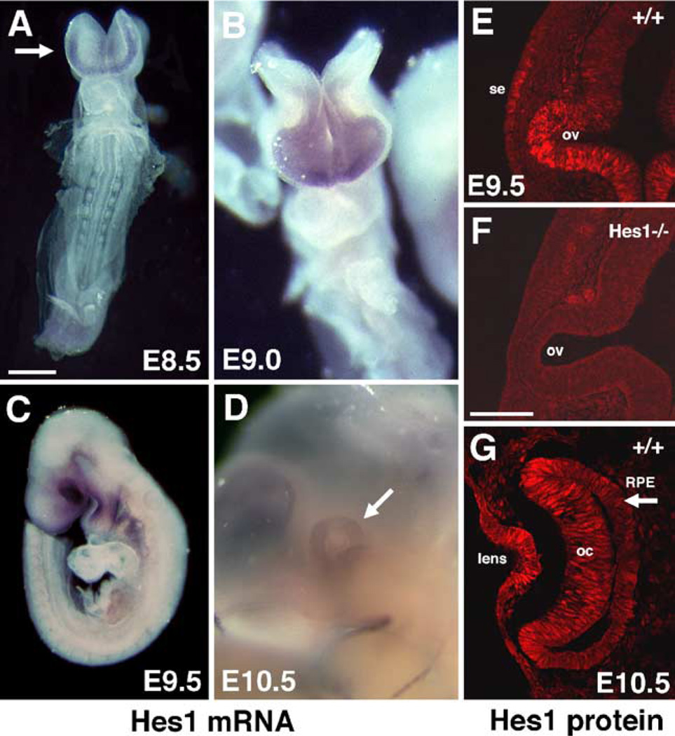
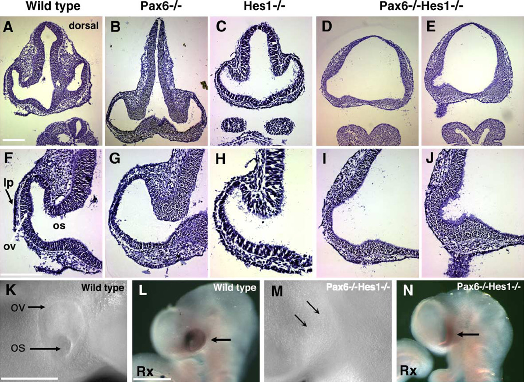
During embryogenesis, multiple developmental processes are integrated through their precise temporal regulation. Hes1 is a transcriptional repressor that regulates the timing of mammalian retinal neurogenesis. However, roles for Hes1 in early eye development have not been well defined. Here, we show that Hes1 is expressed in the forming lens, optic vesicle, cup, and pigmented epithelium and is necessary for proper growth, morphogenesis, and differentiation of these tissues. Because Hes1 is required throughout the eye, we investigated its interaction with Pax6. Hes1-Pax6 double mutant embryos are eyeless suggesting these genes are coordinately required for initial morphogenesis and outgrowth of the optic vesicle. In Hes1 mutants, Math5 expression is precocious along with retinal ganglion cell, amacrine, and horizontal neuron formation. In contrast to apparent cooperativity between Pax6 and Hes1 during morphogenesis, each gene regulates Math5 and RGC genesis independently. Together, these studies demonstrate that Hes1, like Pax6, simultaneously regulates multiple developmental processes during optic development.


Figures

Fig. 1
Fig. 1
Hes1 is expressed at the earliest stages of eye development. (A–D) In situ hybridization for Hes1 mRNA. Dorsal is up or upper left. (A, B) Frontal views. Hes1 mRNA is expressed in the E8.5 anterior neural plate (arrow in A) and optic vesicles of E9.0 embryos (B). (C, D) Lateral embryo views with rostral to the left. Hes1 mRNA is strongly expressed in the E9.5 optic region (C). At E10.5, Hes1 mRNA persists throughout the optic cup (arrow in D). (E–G) Immunofluorescent antibody labeling with anti-Hes1 antibody. (E, F) Cryosections through E9.5 wild type (E) and Hes1−/− (F) optic vesicles. Hes1 protein expression in both surface ectoderm (se) and optic vesicle (ov) are missing in mutants (F). (G) At E10.5, Hes1 protein expression is apparent in the lens, optic cup (oc), and RPE (arrow). Scale bar = 500 µm in panel A, 20 µm in panel F.
Fig. 2
Fig. 2
Early specification and patterning are normal in Hes1 mutants. Wild type and Hes1 mutant embryos labeled with Pax2, Chx10, and Mitf antibodies or in situ hybridization using Rx, Mitf, Dct, and Tyrp1 cRNA probes. In the horizontal sections A–C and E–G, rostral is right and lateral up; in sections D, H–P, rostral is left and dorsal up. (A, E) Pax2 expression (red) in the E9.5 optic vesicle is unaffected in Hes1 mutant embryos. Sections were also counterstained with DAPI (blue). (B, F) Chx10 (green) and Mitf (red) double labeling at E9.5 demonstrates that retina versus RPE/optic stalk boundaries are unaffected in Hes1 mutants. (C, G) In E10.5 wild type, morphogenesis of the optic cup and an almost single cell layered RPE are visible (C). In Hes1 mutants, morphogenesis is arrested at the vesicle stage although retina versus RPE specification is maintained. A few Hes1 mutants had small numbers of cells co-expressing Chx10 and Mitf (yellow domain in between green and red cells in panel G. (D, H) In E10.5 Hes1−/− eyes, Rx mRNA is expressed throughout the future retina as it is in wild type. (I, J, M, N) Mitf mRNA expression in the specified RPE is greatly reduced in E10.5 Hes1−/− embryos (arrow in N). (K, O) Two markers of differentiating RPE, Dct, and Tyrp1 (not shown) are also reduced in Hes1 mutants at E10.5. (L, P) At E12.5, Dct (not shown) and Tyrp1 are clearly expressed but their expression domains smaller than normal in Hes1 mutants. Scale bar = 20 µm in panels A–C, F, G, J, and N, and 500 µm in panels D, H, I, K–M, O, and P.
Fig. 3
Fig. 3
Loss of Hes1 reduces lens and RPE/OS proliferation. Anti-BrdU labeling (in red) of sections containing E9.5 and E10.5 optic regions. White lines in all panels denote tissue boundaries assigned for cell counting. (A, C, E) Representative sections from E9.5 wild type, Hes1+/−, and Hes1−/− eyes showing BrdU incorporation. (B, D, F) Although the E10.5 Hes1−/− eye (F) arrested at the optic vesicle stage, numerous proliferating cells are present. The two shades of red among the sets of images are due to pseudo coloring by different computer imaging programs. (G) The number of cells in S-phase (BrdU antibody labeling) was divided by the total number of nuclei (DAPI labeling, not shown) to give a labeling index (% BrdU labeled cells) on the y axis plotted versus developmental age and tissue type (x axis) for wild type, Hes1+/−, and Hes1−/− embryos. Statistically significant changes for the E9.5 RPE/OS and E10.5 lens are denoted by a bracket and the P values obtained using ANOVA and Fisher’s test. The black line on each bar of the graph represents standard error of the mean. lp = lens placode, pr = presumptive retina, rpe = retinal pigmented epithelium, rpe/os = region of the optic vesicle that gives rise to both structures. Scale bar = 20 µm in panel A.
Fig. 4
Fig. 4
Math5, Ngn2, and Mash1 are selectively derepressed in Hes1 mutants. β-galactosidase or mRNA expression in wild type and Hes1−/− optic vesicles (E9.5) and cups (E10.5–11.5), examined in pairs of images of whole mounts and sections. In panels A, C, E, G, I, K, M, and O, nasal is left and dorsal up and white arrows point to the eye. In panels B, D, F, H, J, L, N, and P, medial is up and rostral is left. (A–D) Comparison of 22-somite Hes1 +/+;Math5LacZk.i./+ and Hes1−/−;Math5lacZk.i./+ littermate embryos at E9.5. No β-gal-positive cells are found in control embryos (A, B). Math5lacZ expression is precocious in Hes1 mutant optic vesicles (C, D). Insets in panels A and C show the optic vesicle at higher magnification. (E–H) Ngn2 is ectopically expressed in the optic stalk at E9.5. Premature expression of Ngn2 in cross section through the forming optic stalk of Hes1 mutants (arrow in H). Precocious Ngn2 expression is not detected in the optic vesicle/cup at E9.5 or E10.5. (I–L) Mash1 mRNA is upregulated in a small dorsal domain at E10.5 in Hes1 mutants. Arrow in panel L denotes a small group of cells inappropriately expressing Mash1. (M–P) At E11.5, Ngn2 mRNA is ectopically expressed in both the optic cup and stalk. Ngn2 expression is upregulated in a Hes1 mutant with a nearly normal optic cup (O). Cross section through an E11.5 Hes1 mutant eye with arrested morphogenesis and ectopic Ngn2 expression in the optic vesicle (thin arrow) and stalk (thick arrow). Scale bar in panel A = 500 µm, panel B = 50 µm.
Fig. 5
Fig. 5
Early retinal neuron cell types have accelerated differentiation in Hes1 mutants. Antibody labeling with anti-βIII-tubulin, an early marker of neuronal differentiation. Wild type optic vesicle (A) and cup (D) sections do not contain neurons at these stages. (A–C) Anti-βIII-tubulin labeling at embryonic day 9.5 (E9.5) demonstrates the presence of a few precocious neurons in Hes1 heterozygotes (arrow in B) but many more in homozygous mutants (C). Differentiating neurons often exhibit neuronal morphology, including neurite extension (arrow in C). (D–F) E10.5 optic cups in wild type, heterozygous, and homozygous Hes1 mutants. Hes1+/− and Hes1−/− optic cups have precocious neurons (arrows), with many more in homozygous mutants. Ectopic neurons in Hes1 mutant are also found in the optic stalk (C) and RPE (arrowheads in F). (G–N) Optic cup sections of E10.5 Hes1−/− embryos, with vitreal to the right. (G) βIII-tubulin (green) and the RGC marker Doublecortin (red) completely overlap (yellow). (H) Co-labeling with the RGC marker NF-160 and βIII-tubulin further indicates that precocious neurons exhibit RGC characteristics. (I) βIII-tubulin+ cells do not express the amacrine cell marker Syntaxin. (J, K) Double-labeling with βIII tubulin and RGC markers Brn3a (J) or Brn3b (K) indicate that the neurons express both markers. (L) βIII-tubulin+ neurons also express Isl1, found in RGCs, and amacrine neurons. (M) Dlx, a marker for early RGCs, horizontal, amacrine, and bipolar neurons in wild type is not expressed in Hes1−/− optic vesicles. (N) Hes1−/− cells expressing either the amacrine/horizontal cell marker VC1.1 alone (red cells) or both βIII-tubulin and VC1.1 (arrow points to the co-labeling in yellow). rpe = retinal pigmented epithelium, nr = neural retina, ov = optic cup, os = optic stalk. Scale bar = 50 µm in panel A, 20 µm in panel G.
Fig. 6
Fig. 6
Hes1 and Pax6 regulation of Math5. Math5 in situ hybridization of E11.0 embryos with differing dosages of Pax6 and Hes1. Panels A, C, E, G, I, and K are whole-mount images of the optic region with nasal left and dorsal up. A white line depicts the plane of section shown in panels B, D, F, H, J, and L horizontal sections from the embryo in each preceding panel, with lateral up. (A, B) Wild type Math5 expression in the dorsal optic cup. (C, D) A Hes1 homozygous mutant with strong Math5 expression. (E, F) In embryos with one mutant copy of Pax6 and Hes1, eyes are smaller than wild type with few Math5-expressing cells. This phenotype is identical to Pax6 heterozygotes (Brown et al., 1998). (G, H) An abnormal optic cup in Pax6+/−;Hes1−/− embryos, where Math5-expressing cells are numerous compared to double heterozygotes. The only difference between eyes in panels E and F, and G and H is increasing loss of Hes1. (I, J) In single Pax6 mutants, all Math5 expression is absent. Note arrested optic vesicle development compared to panels A–H. (K, L) Embryos mutant for both Pax6 and Hes1 completely lack an eye. In serial sections, only a slight evagination of the diencephalon could be found (arrows). L = lens, ov = optic vesicle, asterisks indicate forming brachial arch in panels A, C, E, G, I, and K. Scale bar = 500 µm in panel A, 50 µm in panel B.
Fig. 7
Fig. 7
Hes1/Pax6 double mutant embryos lack optic vesicles. (A–J) Hematoxylin-stained 10-µm frontal sections through the optic region of E9.5 embryos. Panels F–J are higher magnifications of the same sections. (A, F) 28-somite wild type embryo sections containing lens placode (lp), optic vesicle (ov), and optic stalk (os). (B, G) 27-somite Pax6−/− embryo that lacks a lens placode and has open optic vesicle morphology (G). (C, H) Hes1−/− 22-somite embryo with a missing lens placode and abnormal optic vesicle shape (H). (D, E, I, J) A Pax6−/−Hes1−/− 24-somite embryo at two different section depths (panels D, I are more rostral than panels E, J). A narrow bulge of the diencephalon neural tube is seen in 1 –2 sections per embryo (J). Otherwise, these embryos lack a recognizable optic vesicle and exhibit abnormal fore- and hindbrain formation (not shown). (K–N) Whole-mount images of live embryos (K, M) or embryos post-Rx in situ hybridization (L, N). Image in panel K is of the embryo sectioned in panels A and F, the image in panel M is of the embryo in panels D, E, I, and J. At the surface ectoderm of the double mutant embryo, arrows point to the position where the optic vesicle and lens placode should be observable. In panel N, a band of cells express Rx (arrow), thus are specified for optic fate. Scale bar = 50 µm in panels A and G, 500 µm in panels K and L.

Similar articles

Cited by

References

    1. Ahmad I, Dooley CM, Polk DL. Delta-1 is a regulator of neurogenesis in the vertebrate retina. Dev. Biol. 1997;185:92–103. - PubMed
    1. Akagi T, Inoue T, Miyoshi G, Bessho Y, Takahashi M, Lee JE, Guillemot F, Kageyama R. Requirement of multiple basic helix-loop-helix genes for retinal neuronal subtype specification. J. Biol. Chem. 2004;279:28492–28498. - PubMed
    1. Alexiades MR, Cepko CL. Subsets of retinal progenitors display temporally regulated and distinct biases in the fates of their progeny. Development. 1997;124:1119–1131. - PubMed
    1. Andreazzoli M, Gestri G, Cremisi F, Casarosa S, Dawid IB, Barsacchi G. Xrx1 controls proliferation and neurogenesis in Xenopus anterior neural plate. Development. 2003;130:5143–5155. - PubMed
    1. Arimatsu Y, Naegele JR, Barnstable CJ. Molecular markers of neuronal subpopulations in layers 4, 5, and 6 of cat primary visual cortex. J. Neurosci. 1987;7:1250–1263. - PMC - PubMed

Publication types

MeSH terms