Comparative Study
. 2005 Jul 21;47(2):191-199.
doi: 10.1016/j.neuron.2005.06.030.

Abeta42 is essential for parenchymal and vascular amyloid deposition in mice

Affiliations
Comparative Study

Abeta42 is essential for parenchymal and vascular amyloid deposition in mice

Eileen McGowan et al. Neuron. .

Abstract

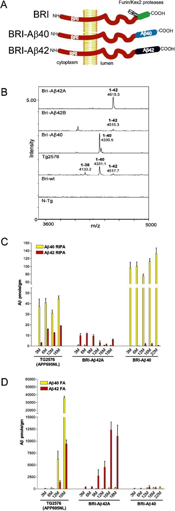
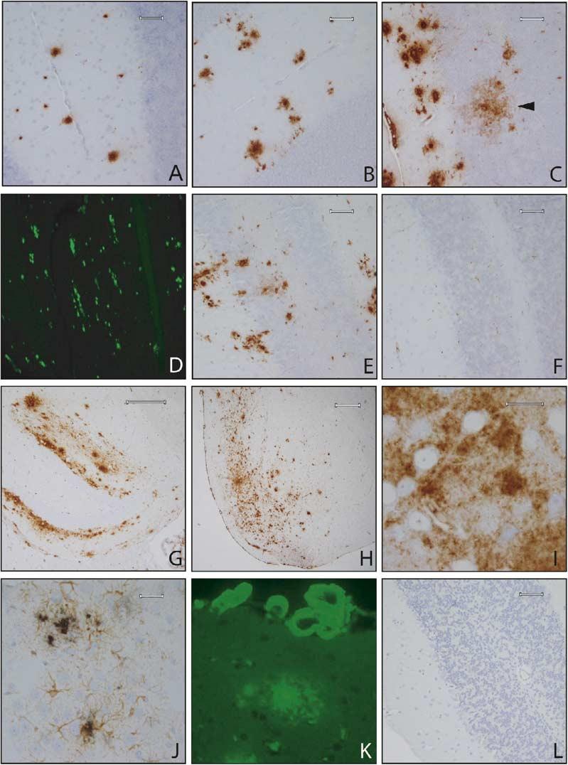
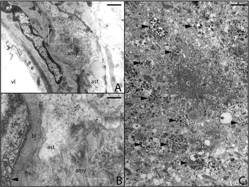
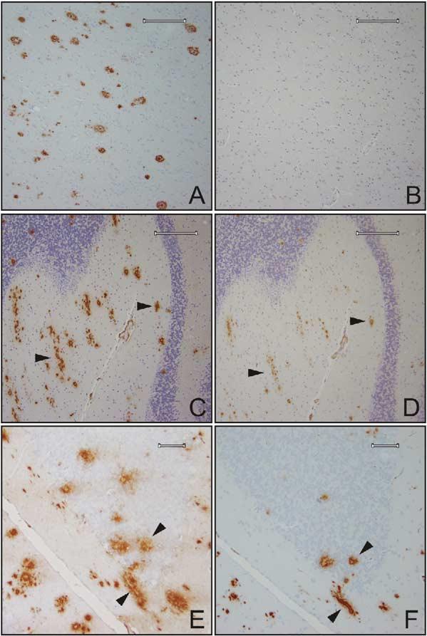
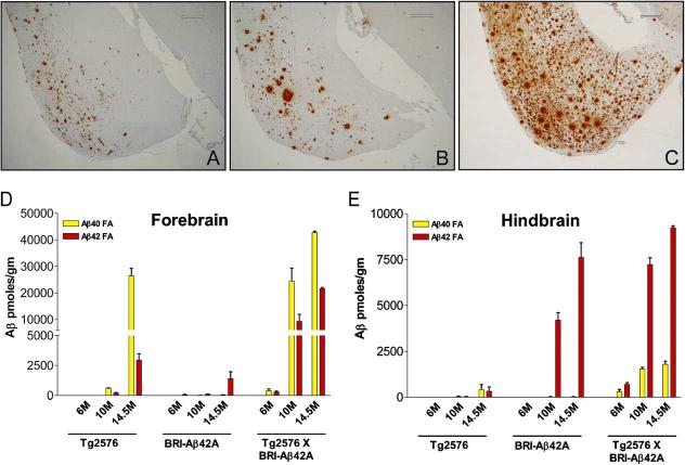
Considerable circumstantial evidence suggests that Abeta42 is the initiating molecule in Alzheimer's disease (AD) pathogenesis. However, the absolute requirement for Abeta42 for amyloid deposition has never been demonstrated in vivo. We have addressed this by developing transgenic models that express Abeta1-40 or Abeta1-42 in the absence of human amyloid beta protein precursor (APP) overexpression. Mice expressing high levels of Abeta1-40 do not develop overt amyloid pathology. In contrast, mice expressing lower levels of Abeta1-42 accumulate insoluble Abeta1-42 and develop compact amyloid plaques, congophilic amyloid angiopathy (CAA), and diffuse Abeta deposits. When mice expressing Abeta1-42 are crossed with mutant APP (Tg2576) mice, there is also a massive increase in amyloid deposition. These data establish that Abeta1-42 is essential for amyloid deposition in the parenchyma and also in vessels.


Figures

Figure 1.
Figure 1.
Biochemical Analyses of Aβ Levels in BRI-Aβ Mice (A) Schematic of the BRI-Aβ fusion proteins. BRI-Aβ fusion constructs were designed to take advantage of the BRI protein that is cleaved by furin or a furin-like protease near the COOH-terminus to release a soluble 23 amino acid peptide in the wild-type protein. Aβ1-40 or Aβ1-42 was fused to the C terminus of the BRI protein at the furin cleavage site. This cleavage releases Aβ into the lumen or extracellular space, resulting in efficient secretion. (B) IP/MS analysis of Aβ isoforms in BRI-Aβ mice. The spectra from the BRI-Aβ42 lines had single peaks detected with a mass/charge ratio (m/z) of 4515 corresponding to human Aβ1-42. In BRI-40 mice, one peak was detected at m/z 4330 corresponding to human Aβ1-40. The shoulder of the 4330 peak corresponds to the Aβ40 sodium adduct ions. Peaks corresponding to human Aβ1-38, Aβ1-40, and Aβ1-42 were detected in extracts from Tg2576 and were absent in nontransgenic mice and BRI wild-type mice. (C) RIPA-soluble (RIPA) and (D) RIPA-insoluble formic acid extractable (FA) Aβ levels in hemibrains (n = 3-7 mice per age point per group) from BRI-Aβ mice were quantified by Aβ sandwich ELISAs. FA Aβ42 increased dramatically with age in the BRI-Aβ42 mice. There was no evidence for accumulation of insoluble Aβ40 in the BRI-Aβ40 mice with age.
Figure 2.
Figure 2.
Amyloid Pathology in BRI-Aβ Mice Progression of amyloid deposition (total Aβ immunostaining with 33.1.1 raised against Aβ1-16) in the cerebellum of BRI-Aβ42A mice at 3 months (A), 6 months (B), and 16.5 months (C). BRI-Aβ42 mice develop both diffuse (arrow in [C]) and compact thioflavin S-positive deposits (D). Plaques were immunoreactive with Aβ42 end-specific antibodies (E). No pathology was evident even in 16.5-month-old BRI-Aβ40 mice, immuno-stained with a panel of Aβ antibodies or thioflavin S (F). Old (19 month) BRI-Aβ42 mice show widespread forebrain Aβ deposits, particularly in the molecular layer of the hippocampus (G) and the entorhinal/piriform cortex (H). The entorhinal Aβ deposits were largely diffuse, and neurons were often observed within these diffuse deposits (I). Increased gliosis was associated with plaque deposition (J). Sections were immunostained with anti-GFAP (brown reaction product) followed by immunostaining for Aβ using the antibody BAM-10 (black reaction product). CAA was progressive and severe in old mice (K); vessels were stained with thioflavin S. A dense cored plaque is also visible. The vascular deposits were fibrillar, as demonstrated by electron microscopy (see Figure 3). (L) wild-type BRI mice had no amyloid pathology (total Aβ immunohistochemistry) at ages up to 24 months of age. Scale bars: 60 μm (A—F), 200 μm (G), 300 μm (H), 20 μm (I), 30 μm (J).
Figure 3.
Figure 3.
Electron Microscopy of Congo-philic Amyloid Angiopathy and Aβ Plaques in BRI-Aβ42 Mice CAA was pronounced in BRI-Aβ42 mice. (A) Amyloid deposit (amy) at the outer basal lamina (marked with *) of an arteriole found at the cortical surface. An interendothelial junction is indicated by the double arrowhead. The boxed area in (A) is enlarged in (B). (B) Note the disruption of basal lamina (bl) between the astrocyte (ast) and smooth muscle cells (smc) by bundles of amyloid fibrils (amy). Arrowhead points to surface vesicles of SMC. Many of the extracellular amyloid plaques in the BRI-Aβ mice had dense amlyoid aggregates with radiating fibrils (C). There were many dystrophic neurites (arrow-heads) present at the periphery of these plaques. vl, lumen; e, endothelium; SMC, smooth muscle cell; Ast, astrocytes. Scale bars: 1 μm (A), 30 nm (B), and 5 μm (C).
Figure 4.
Figure 4.
Mouse Aβ and Modified Aβ Peptides Are Present in the Plaques of BRI-Aβ42 Mice (A) Senile plaques in the temporal gyrus from an AD brain, Braak stage VI, immunostained with anti-Aβ1-16 (33.1.1). (B) A serial section of the AD tissue in (A) immunostained with anti-rodent Aβ that does not detect human Aβ deposited in AD. (C) Total Aβ immunostaining using antibody 33.1.1, in the cerebellum of a 17-month-old BRI-Aβ mouse shows multiple cored plaques, a subset of these plaque cores are immunopositive for rodent Aβ in a serial section (D). Arrows in (C) and (D) indicate the same plaques immunostained with 33.1.1 and rodent Aβ, respectively. (E) Diffuse and dense cored plaques in the cerebellum were immunopositive with an anti-Aβ1-16 (82E1, IBL), a subset of these plaques were stained by an antibody that recognizes a truncated N-terminal modified pyrogluta-mate form of Aβ (N3pE Aβ) (F). Arrows in (E) and (F) indicate the same plaques immunostained with anti-Aβ1-16 and anti-N3pE Aβ, respectively. Scale bars: 200 μm (A—D) and 60 μm (E and F).
Figure 5.
Figure 5.
Enhanced Pathology in BRI-Aβ42 × Tg2576 Bigenic Mice Transgenic mice expressing both BRI-Aβ42 and mutant APPswe(Tg2576) have enhanced senile plaque pathology in the forebrain compared with their singly transgenic littermates and significantly elevated insoluble Aβ levels in the forebrain and hindbrain. Entorhinal/piriform cortex of 14.5-month-old BRI-Aβ42A mouse (A), 14.5-month-old Tg2576 (B), 14.5-month-old BRI-Aβ42 × Tg2576 bigenic mouse (C) immunostained with anti-total Aβ (33.1.1). All mice were littermates. FA Aβ levels in the forebrain (D) and hindbrain (E) of an aging series of bigenic BRI-Aβ/Tg2576 compared with their single transgenic littermates. Scale bars: 300 μm (A—C).

References

    1. Bales KR, Verina T, Cummins DJ, Du Y, Dodel RC, Saura J, Fishman CE, DeLong CA, Piccardo P, Petegnief V, et al. Proc. Natl. Acad. Sci. USA. Vol. 96. 1999. Apolipoprotein E is essential for amyloid deposition in the APP(V717F) transgenic mouse model of Alzheimer's disease; pp. 15233–15238. - PMC - PubMed
    1. Borchelt DR, Davis J, Fischer M, Lee MK, Slunt HH, Ratovitsky T, Regard J, Copeland NG, Jenkins NA, Sisodia SS, Price DL. A vector for expressing foreign genes in the brains and hearts of transgenic mice. Genet. Anal. 1996;13:159–163. - PubMed
    1. Borchelt DR, Ratovitski T, van Lare J, Lee MK, Gonzales V, Jenkins NA, Copeland NG, Price DL, Sisodia SS. Accelerated amyloid deposition in the brains of transgenic mice coexpressing mutant presenilin 1 and amyloid precursor proteins. Neuron. 1997;19:939–945. - PubMed
    1. Chishti MA, Yang DS, Janus C, Phinney AL, Horne P, Pearson J, Strome R, Zuker N, Loukides J, French J, et al. Early-onset amyloid deposition and cognitive deficits in transgenic mice expressing a double mutant form of amyloid precursor protein 695. J. Biol. Chem. 2001;276:21562–21570. - PubMed
    1. Farris W, Mansourian S, Chang Y, Lindsley L, Eckman EA, Frosch MP, Eckman CB, Tanzi RE, Selkoe DJ, Guenette S. Insulin-degrading enzyme regulates the levels of insulin, amyloid beta-protein, and the beta-amyloid precursor protein intra-cellular domain in vivo. Proc. Natl. Acad. Sci. USA. 2003;100:4162–4167. - PMC - PubMed

Publication types

MeSH terms

LinkOut - more resources