. 2006 Jan 15;150(1):80-9.
doi: 10.1016/j.jneumeth.2005.06.002. Epub 2005 Jul 21.

Reflexes evoked by electrical stimulation of afferent axons in the pudendal nerve under empty and distended bladder conditions in urethane-anesthetized rats

Affiliations

Reflexes evoked by electrical stimulation of afferent axons in the pudendal nerve under empty and distended bladder conditions in urethane-anesthetized rats

Hui-Yi Chang et al. J Neurosci Methods. .

Abstract

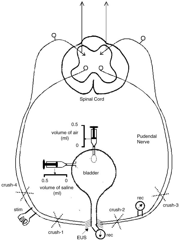
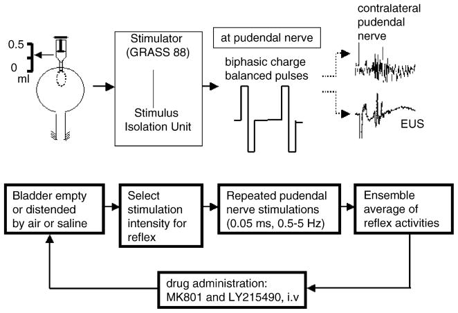
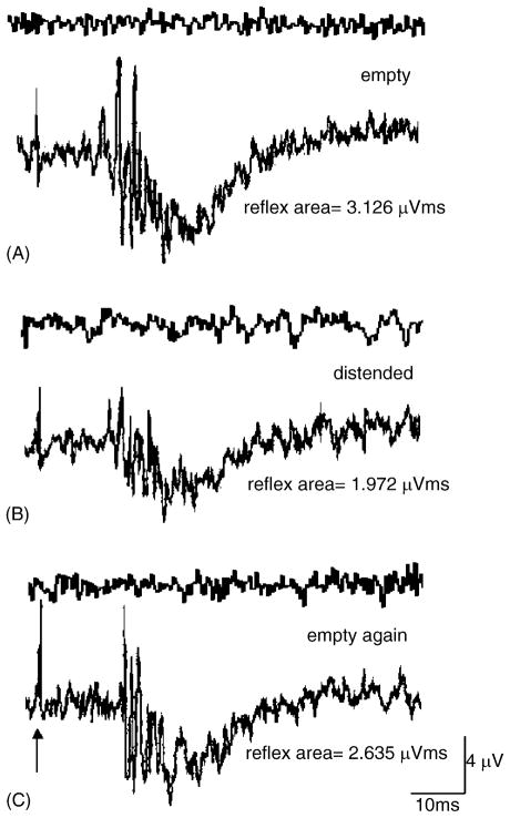
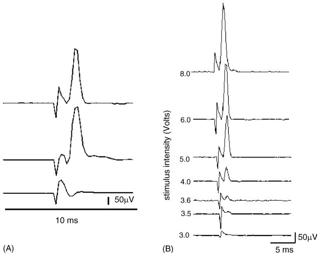
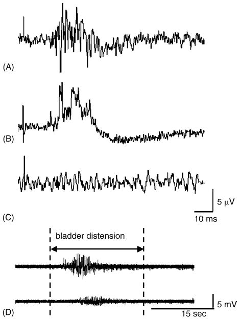
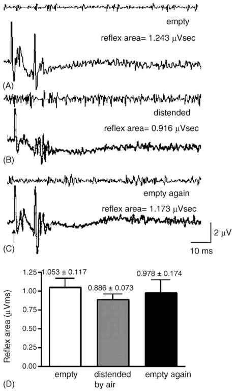
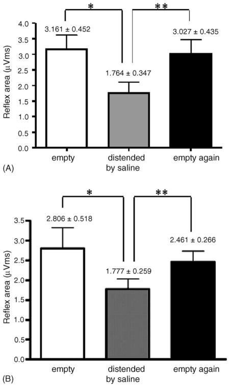
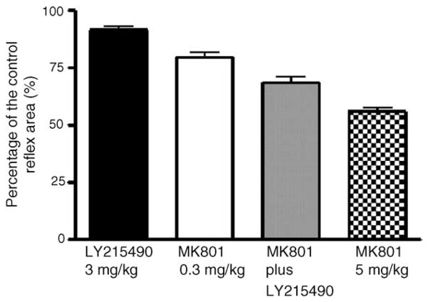
This study examined reflex mechanisms that mediate urinary bladder and external urethral sphincter (EUS) coordination in female Sprague-Dawley urethane-anesthetized rats under empty and distended bladder conditions. The bladder was distended either by a small balloon or a saline filled catheter inserted through the body of the bladder. Stimulation of the entire pudendal nerve elicited short latency (8-12 ms) responses in the EUS and short (3-8 ms) and long latency responses (16-20 ms) in contralateral pudendal nerve. The long latency pudendal-pudendal reflex was reduced by 36.7% in area during bladder distension with the balloon catheter. However, there was no significant change in the area of pudendal-EUS reflex during bladder distension. Peak amplitudes of both reflexes were reduced 32% by bladder distension. The effects of glutamatergic receptor antagonists on the reflexes were also examined. MK 801 (0.3-5mg/kg, i.v.), an N-methyl-d-aspartate glutamatergic receptor antagonist, markedly depressed the pudendal-pudendal reflex, but LY 215490 (3mg/kg, i.v.), an alpha-amino-5-methyl isoxazole-4-propionate antagonist, had a minimal inhibitory effect. Both glutamatergic receptor antagonists significantly suppressed the pudendal-EUS reflex. These results indicate that the EUS is innervated by multiple pathways and that glutamatergic excitatory transmission is important in the neural mechanisms underlying bladder-sphincter coordination in the rat.


Figures

Fig. 1
Fig. 1
Schematic diagram of the experimental setup. Stimulation electrodes (stim) were positioned on the pudendal nerve on one side and recording electrodes (rec) were positioned on the nerve on the contralateral side approximately 35 mm from the junction of the nerves with the urethra. During the experiments the pudendal nerves were crushed peripheral (#1 and #2) or central (#3 and #4) to the electrodes to determine the pathways for evoking the reflexes. The EUS–EMG was recorded by inserting single electrodes into the striated muscle on either side of the urethra. Catheters were inserted into the bladder lumen to distend the bladder. One catheter was connected to a small balloon that could be filled with air; the other catheter was filled with saline.
Fig. 2
Fig. 2
Flowchart of the experimental protocol for the electrophysiological and pharmacological studies. Reflex activity in pudendal efferent nerves and EUS–EMG were elicited by electrical stimulation of pudendal afferent axons using biphasic charge balanced pulses. The bladder was distended with air or saline. After completing the physiological studies reflexes were recorded before and after injection of glutamatergic receptor antagonists administered as single doses to individual animals or administered sequentially, MK801 followed by LY215490. Recordings were obtained 25–45 min after drug injection.
Fig. 3
Fig. 3
Measurement of the magnitude of the pudendal nerve reflex. Reflex area represents area (μV ms) under the upward deflection of the evoked potential between two cursors set on the baseline at the beginning and the end of the potential. Usually the potential consisted of several peaks of varying amplitude. Amplitude measurements reflect the amplitude of the largest peak (μV).
Fig. 4
Fig. 4
Effect of bladder distension on the pudendal nerve reflex elicited by a single shock to the contralateral pudendal nerve (arrow) (A) when the bladder was empty, (B) when the bladder was distended with 0.2 ml of saline, and (C) after recovery following bladder distension. Upper trace in each pair of recordings is the control activity in the absence of stimulation. Lower trace shows the response to stimulation.
Fig. 5
Fig. 5
EUS–pudendal response. (A) Upper trace: the pudendal nerve response (average of 10 individual responses) was elicited by electrical stimulation of the EUS (6 V, 0.7 Hz, pulse width 0.05 ms). Middle trace: the EUS–pudendal response was not changed after the contralateral pudendal nerve was crushed at the site marked crush-1 in Fig. 1. Lower trace: the pudendal response was totally abolished after the pudendal nerve was crushed at the site marked crush-2 in Fig. 1. (B) Changes in the EUS–pudendal response with stimulus intensity. Series of recordings showing the EUS–pudendal response at increasing stimulus intensities. The threshold was identified as 3.5 V in this animal. Only the stimulation artifact occurs below the threshold (3.0 V, the lowest trace). The first trace shows the maximum amplitude of this response elicited by 8 V. Each trace represents the average of 10 individual responses. The stimulus duration was constant at 0.05 ms.
Fig. 6
Fig. 6
Effect of crushing the pudendal nerves at various sites on the pudendal–pudendal (A) and bladder–EUS reflexes. (A) The pudendal–pudendal reflex was elicited by single-shock stimulation (9 V, 0.5 Hz, 0.05 ms pulse duration) when the bladder was distended. (B) The pudendal–pudendal reflex remained after the pudendal nerve was crushed at the site marked crush-2 in Fig. 1. (C) The pudendal–pudendal reflex was abolished after pudendal nerves were crushed bilaterally at sites marked crush-3 and crush-4 in Fig. 1. (D) After bilateral crushing of pudendal nerves, the EUS–EMG activity induced by bladder distension was reduced but not completely eliminated. Upper panel: EUS–EMG firing induced by distending the bladder with saline when the pudendal nerves were intact. Lower panel: after crushing pudendal nerves bilaterally the amplitude of EUS–EMG activity during bladder distension was markedly reduced.
Fig. 7
Fig. 7
Examples of the pudendal–EUS reflex when the bladder was (A) empty, (B) distended by air, and (C) empty after distension. (D) Statistical analysis indicated no significant change in the reflex area of the pudendal–EUS reflex when the bladder was distended by air or empty (N = 6). However the amplitude of the reflex was reduced during bladder distension (B).
Fig. 8
Fig. 8
Effect of bladder distension with different volumes of saline and air on the pudendal–pudendal reflex (A). The reflex area of the pudendal–pudendal reflex was compared when the bladder was empty, distended by saline (0.2 ml) and emptied again. Statistical analysis indicated that the pudendal–pudendal reflex was significantly decreased by distension of the urinary bladder (N = 6, P < 0.05, Student’s t-test). (B) Similarly, the reflex area was significantly changed when the bladder was distended by 0.5 ml of air (*). The reflex area was significantly increased after the bladder was emptied (**). There was no significant difference between control and empty conditions.
Fig. 9
Fig. 9
The effects MK801 and LY2155490 on the reflex area of the pudendal–pudendal reflex (N = 12). The animals were separated into three groups: group 1 (N = 3) receiving LY215490 alone (3.0 mg/kg, i.v.), group 2 (N = 6) first receiving MK801 (0.3 mg/kg, i.v.) and followed by LY215490 (3.0 mg/kg, i.v.), and group 3 (N = 3) receiving MK801 alone (5.0 mg/kg, i.v.). The reflex area was decreased by an average of 23% with MK801 (0.3 mg/kg, i.v.), and 45% with MK801 (5.0 mg/kg, i.v.). When LY215490 (3.0 mg/kg, i.v.), was administered after MK801 (0.3 mg/kg, i.v.) the reflex area exhibited only a small further reduction (4%). LY215490 (3.0 mg/kg, i.v.) alone had a small suppressant effect (9%).

References

    1. Boggs JW, Wenzel BJ, Gustafson KJ, Grill WM. Spinal micturition reflex mediated by afferents in the deep perineal nerve. J Neurophysiol. 2005;93:2688–97. - PubMed
    1. de Groat WC. Anatomy and physiology of the lower urinary tract. Urol Clin North Am. 1993;20:383–401. - PubMed
    1. de Groat WC, Booth AM, Yoshimura N. Neurophysiology of micturition and its modification in animal models of humane disease. In: Maggi CA, editor. The autonomic nervous system: nervous control of the urogenital system. Vol. 3. London, UK: Harwood Academic Publishers; 1993. pp. 227–89.
    1. Floyd K, McMahon SB, Morrison JFB. Inhibitory interactions between colonic and vesical afferents in the micturition reflex of the cat. J Physiol. 1982;322:45–52. - PMC - PubMed
    1. Jezernik S, Craggs M, Grill WM, Creasey G, Rijkhoff NJ. Electrical stimulation for the treatment of bladder dysfunction: current status and future possibilities. Neurol Res. 2002;24:413–30. - PubMed

Publication types

MeSH terms

LinkOut - more resources