. 2005 Aug;138(4):1842-52.
doi: 10.1104/pp.105.063537. Epub 2005 Jul 22.

An RNA-dependent RNA polymerase prevents meristem invasion by potato virus X and is required for the activity but not the production of a systemic silencing signal

Affiliations

An RNA-dependent RNA polymerase prevents meristem invasion by potato virus X and is required for the activity but not the production of a systemic silencing signal

Frank Schwach et al. Plant Physiol. 2005 Aug.

Abstract

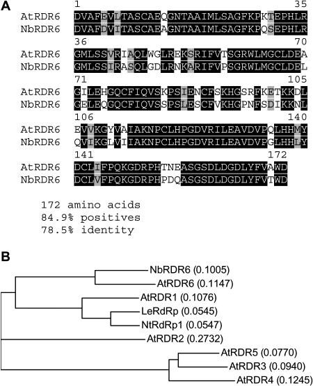
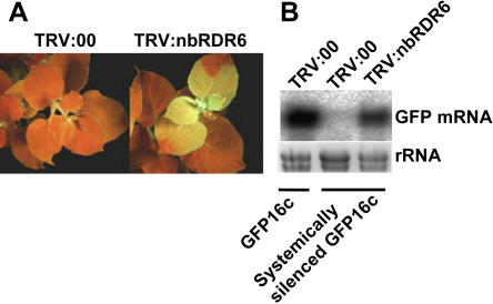
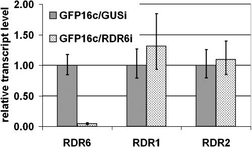
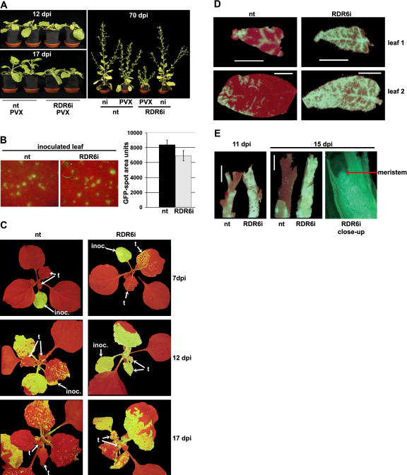
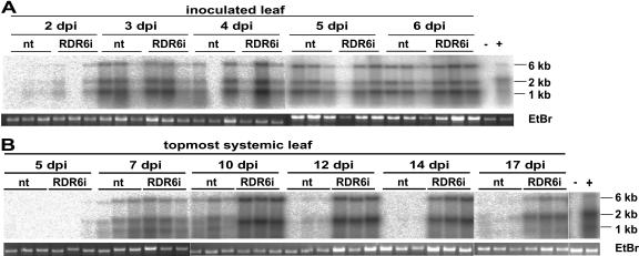
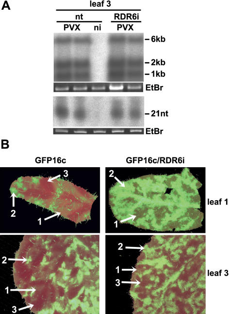
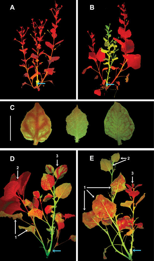
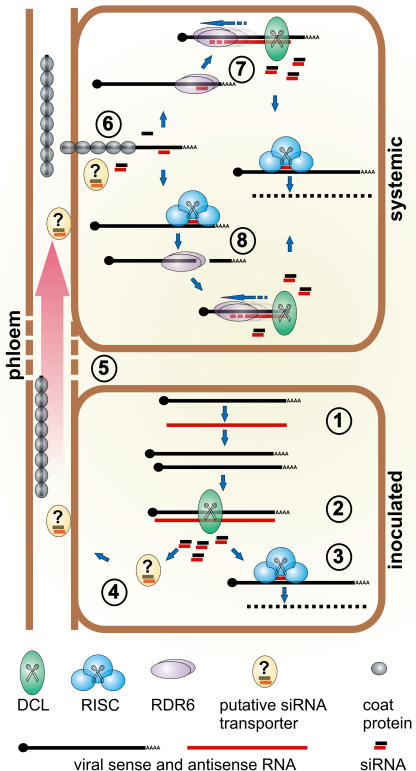
One of the functions of RNA silencing in plants is antiviral defense. A hallmark of RNA silencing is spreading of the silenced state through the plant. Little is known about the nature of the systemic silencing signal and the proteins required for its production, transport, and reception in plant tissues. Here, we show that the RNA-dependent RNA polymerase RDR6 in Nicotiana benthamiana is involved in defense against potato virus X at the level of systemic spreading and in exclusion of the virus from the apical growing point. It has no effect on primary replication and cell-to-cell movement of the virus and does not contribute significantly to the formation of virus-derived small interfering (si) RNA in a fully established potato virus X infection. In grafting experiments, the RDR6 homolog was required for the ability of a cell to respond to, but not to produce or translocate, the systemic silencing signal. Taking these findings together, we suggest a model of virus defense in which RDR6 uses incoming silencing signal to generate double-stranded RNA precursors of secondary siRNA. According to this idea, the secondary siRNAs mediate RNA silencing as an immediate response that slows down the systemic spreading of the virus into the growing point and newly emerging leaves.


Figures

Figure 1.
Figure 1.
Alignment of the translated NbRDR6 sequence fragment to Arabidopsis RDR6 (A) and guide tree (B) of translated NbRDR6 aligned with all six Arabidopsis RDRs (AtRDR1AtRDR6) and with RDR proteins from tomato and tobacco (LeRdRP and NtRdRP1, respectively). Calculated distance values according to the Neighbor Joining method (Saitou and Nei, 1987) are given in parentheses.
Figure 2.
Figure 2.
Virus-induced gene silencing of NbRDR6 in N. benthamiana. Two-week-old GFP16c plants were systemically silenced for GFP by transient expression of a GFPi. Once silencing was established, TRV VIGS vectors either containing the NbRDR6 fragment (TRV:RDR6) or empty (TRV:00) were inoculated. Five to six weeks later, TRV:RDR6-inoculated but not TRV:00-inoculated plants showed a breakdown of maintenance of the GFP silencing, visible as green fluorescence under UV light (A) and reestablished expression of the GFP mRNA (B) in the virus-infected tissues.
Figure 3.
Figure 3.
Quantitative real-time RT-PCR for RDR6, RDR1, and RDR2 transcript levels in N. benthamiana lines carrying the RDR6i construct or an unrelated RNAi construct (GUSi). Mean values are based on four pools of five plants each. Error bars represent ses of the mean. Relative transcript levels were calculated using the ΔΔC(t) method (Livak and Schmittgen, 2001) with GAPDH transcripts serving as an internal standard.
Figure 4.
Figure 4.
Comparison of PVX:GFP infection on N. benthamiana nt and RDR6i lines. A, RDR6i and nt plants were inoculated with PVX:GFP and are shown at 12 dpi when there was no difference in appearance of the two plant lines and at 17 dpi when the RDR6i plants were stunted. At 70 dpi, the RDR6i line still exhibited strong symptoms, whereas the nt plants exhibited mild symptoms and were similar to noninoculated plants. B, PVX:GFP infection foci in inoculated leaves of nt and RDR6i plants at 4 dpi imaged under UV light were similar in size and brightness, indicating that replication and cell-to-cell spread of the virus were unaffected by the RDR6i genotype. Spot sizes were measured using the Able Image Analyser software (Mu Labs, Slovenia) and mean values of area units of 55 spots on each plant line at 4 dpi are shown with their ses of the mean. C, PVX:GFP-infected RDR6i and nt plants under UV light. The inoculated leaves indicated by arrows (inoc.). The two topmost leaves (t) showed spread of PVX:GFP into the upper leaves from 7 dpi, whereas in nt plants the uppermost leaves were largely free of GFP fluorescence up to 17 dpi and after. D, Young leaves (leaf 1 and leaf 2 from top) of PVX:GFP-infected nt and RDR6i plants under UV illumination illustrating a difference in PVX:GFP distribution at 12 dpi. White bars = 0.5 cm. E, Stem tips of nt and RDR6i plants infected with PVX:GFP show more extensive viral spread into young tissue in RDR6i than in nt. At 11 dpi, the top 0.5 cm and at 15 dpi the top 1.5 cm of the nt stem appear red, whereas the RDR6i stem exhibits virus-expressed GFP throughout. A longitudinal section of the growing tip of the RDR6i plant (15 dpi, right-hand section) shows GFP fluorescence evenly distributed throughout, including the meristem.
Figure 5.
Figure 5.
Northern-blot analyses of PVX:GFP RNA levels in nt and RDR6i plants. A, PVX:GFP RNA accumulation in inoculated leaves in nt and RDR6i plants in triplicate samples from 2 to 6 dpi. Accumulation of PVX:GFP full-length (6 kb) and major subgenomic RNAs (approximately 1 and 2 kb) were detected with a DNA probe specific for the PVX coat protein sequence. No significant difference in PVX:GFP RNA accumulation apart from plant-to-plant variation was visible. B, PVX:GFP RNA accumulation in the topmost, not yet fully expanded, leaves in triplicate samples from nt and RDR6i plants at the indicated time points from 5 dpi to 17 dpi is showing increasing differences in viral accumulation between the two plant lines throughout the experiment in the newly developing tissue.
Figure 6.
Figure 6.
Silencing signal and siRNA production in the presence or absence of NbRDR6 in PVX:GFP-infected plants at 10 dpi. A, Northern-blot analysis of PVX:GFP viral genomic (6 kb) and subgenomic (approximately 1 and 2 kb) RNA and accumulation of PVX:GFP-derived siRNA (21 nt) in fully systemically infected leaves (leaf 3 from the top) shows no difference in viral RNA and siRNA accumulation between nt and RDR6i plants once the infection is fully established. DNA or RNA probes specific for the PVX coat protein sequence were used to detect viral full-length and subgenomic RNAs or virus-derived siRNA, respectively. B, Infection of GFP16c and GFP16c/RDR6i with PVX:GFP for simultaneous monitoring of virus spread, GFP silencing, and transgene-derived GFP expression. Images of first and third leaves (from top) were taken under UV light at 10 dpi. Three levels of GFP fluorescence were discernible: background GFP expression from the GFP transgene (1), bright green areas due to GFP expressed by the replicating virus (2), and dark red areas of GFP silenced tissue (3). In the uppermost leaf 1 of GFP16c, the GFP silencing was evident around the veins, whereas replicating virus produced bright green GFP spots superimposed on the faint fluoresce from the GFP transgene. The same patterns were evident in leaf 3 of GFP16c. However, in GFP16c/RDR6i, the virus-derived GFP was more prevalent and there was only limited evidence of silencing adjacent to the infected areas in the older leaf 3, which was not centered around the veins. A magnified version of the green channel only of this image along with a schematic representation of the areas of different GFP expression levels is available as supplementary data (Supplemental Fig. 3).
Figure 7.
Figure 7.
Grafting experiments, RDR6i plants as either receptors or producers of a systemic silencing signal. Graft junctions are indicated by blue arrows and all plants are shown under UV illumination. A, A GFP16c plant grafted as scion onto a GFP16c/GFPi stock exhibited complete silencing of GFP within 2 weeks. B, A GFP16c/RDR6i plant grafted as scion onto a GFP16c/GFPi stock remained unsilenced as shown here after 3 weeks and later. C, Young, not yet fully expanded leaves of GFP16c scions (left) showed GFP silencing around the veins as shown here after 1 week that eventually spread so that the whole leaf appeared red under UV light. Leaves of the same stage of GFP16c/RDR6i scions normally did not exhibit any sign of GFP silencing (middle) but in 6/24 plants a pattern of vein-centered silencing was observed that failed to spread into the rest of the leaf (right). White bar = 0.5 cm. D, Transient expression of GFPi in leaves (1) of a GFP16c rootstock induced systemic silencing in the rootstock (2) and a GFP16c scion (3) at 24 d. E, GFP16c/RDR6i rootstocks exhibited local silencing in inoculated leaves, transiently expressing the GFPi construct (1) but no spread of silencing into newly emerging leaves occurred (2). A silencing signal was still produced in these rootstocks, which induced systemic silencing in GFP16c scions after 24 d (3).
Figure 8.
Figure 8.
Model for RDR6 function in antiviral defense against PVX. In the inoculated cell (lower box) PVX replicates, forming dsRNA with genomic or subgenomic and antisense RNA in the process (1). DCL recognizes the dsRNA as a substrate and produces virus-derived siRNA (2), which guides RISC to the viral RNA (3). siRNA, probably bound to a transporter protein (4), are translocated through the phloem stream into upper parts of the plant alongside the coated viral RNA (5). Viral RNA and virus-derived siRNA are unloaded simultaneously into cells in the upper part of the plant and the virus-derived siRNA can anneal to the unpacked viral RNA immediately (6). Annealed siRNA could either be used as a primer by RDR6 to produce the dsRNA substrate for DCL (7) or they could guide RISC to cleave the viral RNA, leaving an aberrant (noncapped or nonpolyadenylated respectively) RNA that is recognized by RDR6 for unprimed synthesis of the dsRNA substrate for DCL (8). Both pathways lead to immediate production of siRNA to target viral RNA for RISC-mediated destruction. If no signal or no RDR6 is present in the receiving tissue, this immediate response is not possible and production of viral siRNA relies on viral replication, giving the virus a head start before the silencing machinery. Once the infection is fully established, the viral replicase produces abundant substrate for DCL and the RDR6 pathway does not contribute significantly to virus-derived siRNA production anymore.

References

    1. Astier-Manifacier S, Cornuet P (1974) RNA-dependent RNA polymerase in Chinese cabbage. Biochim Biophys Acta 232: 484–493 - PubMed
    1. Baulcombe D (2004) RNA silencing in plants. Nature 431: 356–363 - PubMed
    1. Baulcombe DC, Chapman SN, Santa Cruz S (1995) Jellyfish green fluorescent protein as a reporter for virus infections. Plant J 7: 1045–1053 - PubMed
    1. Beclin C, Boutet S, Waterhouse P, Vaucheret H (2002) A branched pathway for transgene-induced RNA silencing in plants. Curr Biol 12: 684–688 - PubMed
    1. Bernstein E, Caudy AA, Hammond SM, Hannon GJ (2001) Role for a bidentate ribonuclease in the initiation step of RNA interference. Nature 409: 363–366 - PubMed

Publication types

Associated data