. 2005 Aug;138(4):1967-81.
doi: 10.1104/pp.105.061291. Epub 2005 Jul 22.

Arabidopsis peroxin 16 coexists at steady state in peroxisomes and endoplasmic reticulum

Affiliations

Arabidopsis peroxin 16 coexists at steady state in peroxisomes and endoplasmic reticulum

Sheetal K Karnik et al. Plant Physiol. 2005 Aug.

Abstract

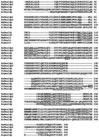
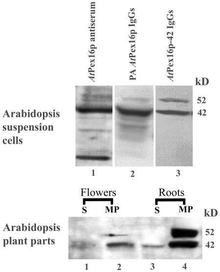
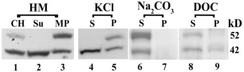
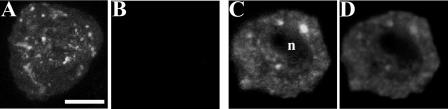
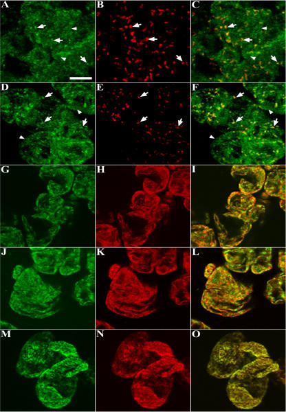
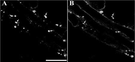
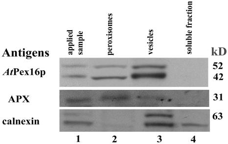
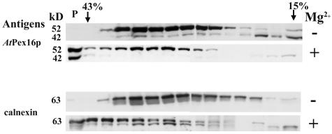
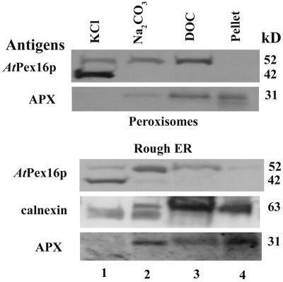
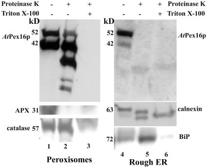
Homologs of peroxin 16 genes (PEX16) have been identified only in Yarrowia lipolytica, humans (Homo sapiens), and Arabidopsis (Arabidopsis thaliana). The Arabidopsis gene (AtPEX16), previously reported as the SSE1 gene, codes for a predicted 42-kD membrane peroxin protein (AtPex16p). Lin et al. (Y. Lin, J.E. Cluette-Brown, H.M. Goodman [2004] Plant Physiol 135: 814-827) reported that SSE1/AtPEX16 was essential for endoplasmic reticulum (ER)-dependent oil and protein body biogenesis in peroxisome-deficient maturing seeds and likely also was involved in peroxisomal biogenesis based on localization of stably expressed green fluorescent protein::AtPex16p in peroxisomes of Arabidopsis plants. In this study with Arabidopsis suspension-cultured cells, combined in vivo and in vitro experiments revealed a novel dual organelle localization and corresponding membrane association/topology of endogenous AtPex16p. Immunofluorescence microscopy with antigen affinity-purified IgGs showed an unambiguous, steady-state coexistence of AtPex16p in suspension cell peroxisomes and ER. AtPex16p also was observed in peroxisomes and ER of root and leaf cells. Cell fractionation experiments surprisingly revealed two immunorelated polypeptides, 42 kD (expected) and 52 kD (unexpected), in homogenates and microsome membrane pellets derived from roots, inflorescence, and suspension cells. Suc-gradient purifications confirmed the presence of both 42-kD and 52-kD polypeptides in isolated peroxisomes (isopycnic separation) and in rough ER vesicles (Mg2+ shifted). They were found peripherally associated with peroxisome and ER membranes but not as covalently bound subunits of AtPex16p. Both were mostly on the matrix side of peroxisomal membranes and unexpectedly mostly on the cytosolic side of ER membranes. In summary, AtPex16p is the only authentic plant peroxin homolog known to coexist at steady state within peroxisomes and ER; these data provide new insights in support of its ER-related, multifunctional roles in organelle biogenesis.


Figures

Figure 1.
Figure 1.
Alignment (ClustalW) of deduced amino acid sequences for Pex16p homologs in humans (Hs; two isoforms), Mus musculus (Mm), Y. lipolytica (Yl), and Arabidopsis (At). Gaps (marked with dashes) were introduced to maximize the sequence alignment. Predicted membrane helices are underlined. Clusters of basic amino acid residues are bolded. Predicted N-linked glycosylation sites are boxed.
Figure 2.
Figure 2.
Rabbit anti-AtPex16p IgGs, affinity purified against Immobilon-bound 42-kD polypeptides (anti-AtPex16p-42 IgGs), are specific for 42- and 52-kD polypeptides in cell fractions of Arabidopsis suspension cells, roots, and leaves. Top, Arabidopsis suspension cell (7 d) homogenates (1,500g, 15 min supernatants from 5,000 psi pressure cell-ruptured cells) were subjected to SDS-PAGE and polypeptides were electroblotted to Immobilon and immunodetected via chemiluminescence with rabbit anti-AtPex16p antiserum (AtPex16p antiserum; 1:1,000, lane 1), PA-purified IgGs (PA anti-AtPex16p IgGs; 1:1,000, lane 2), and 42-kD polypeptide affinity-purified IgGs (anti-AtPex16p-42 IgGs; 1:100, lane 3). Bottom, Roots and flowers excised from 4-week-old Arabidopsis plants were powdered in liquid nitrogen, and then homogenized with a mortar and pestle in a buffered medium. Membrane-free supernatants (S, 200,000g, 1 h; lanes 1 and 3) and corresponding microsomal membrane pellets (MP; lanes 2 and 4), prepared from clarified homogenates, were subjected to SDS-PAGE immunoblot analyses. Samples were detergent (DOC) solubilized, TCA precipitated, and 150 μg protein was applied per well. Chemiluminescence signals were produced from polypeptide-bound anti-AtPex16p-42 IgGs (1:100, overnight incubation).
Figure 3.
Figure 3.
AtPex16p-42 and 52-kD polypeptides are variously associated with Arabidopsis suspension cell microsomal membranes. Clarified homogenates (CH) prepared in HEPES-buffered homogenizing medium (HM; 1,500g, 15 min supernatant of 5,000 psi pressure cell-ruptured cells; lane 1) were centrifuged (200,000g, 1 h) to produce a membrane-free supernatant (Su; lane 2) and microsomal membrane pellet (MP; lane 3). Equal portions of separate water-insoluble microsomal pellets (4 mg protein per 2 mL) were incubated with 0.2 m KCl, 0.1 m Na2CO3 (pH 11.5), or 0.05% (w/v) DOC for 1 h. Supernatant (S) and pellet (P) fractions (200,000g, 30 min) were recovered and analyzed from each treatment (lanes 4–9). These fractions and those from HM were subjected to SDS-PAGE immunoblot analyses (as for Fig. 2) using anti-AtPex16p-42 IgGs (1:100, overnight) as probes for all samples. Protein samples (150 μg) were added per well of SDS gels.
Figure 4.
Figure 4.
PA anti-AtPex16p IgGs specifically recognize myc-epitope-tagged AtPex16p transiently expressed in Arabidopsis suspension cells processed for immunofluorescence microscopy via our standard tube procedure. Arabidopsis suspension cells, biolistically bombarded with pRTL2/mycAtPEX16 and held for 2.5 h expression, were formaldehyde fixed, cell walls permeabilized and digested with pectolyase and cellulase, membranes permeabilized with Triton X-100, and organelles immunolabeled with primary and secondary antibodies in a tube prior to spreading these cells on microscope slides for examination of the transformed immunofluoescent cells (about 0.2% of the cells on the slides). A to D, All images are representative confocal laser optical sections. A and B, Results of dual labeling a representative cell transiently expressing mycAtPex16p. A, Cell illustrating a reticular (ER) and punctate (peroxisome) Cy2 immunofluorescence image labeled with anti-mouse myc epitope antibodies (1: 500, 1 h) and B, the same cell examined for Cy5 bound to anti-AtPex16p-42 IgGs (1:10, overnight). No Cy5 fluorescence attributable to endogenous AtPex16p was observed in the transformed or neighboring nontransformed cells. C and D, Fluorescence images of a cell from the same population of bombarded cells dual labeled with C, anti-mouse myc epitope antibodies (1:500, 1 h), and D, rabbit PA anti-AtPex16p IgGs (1:1,000, 2 h). Colocalization is apparent between the myc (Cy2) and AtPex16p (Cy5) labeled antigens in both reticulate and punctate patterns observed throughout the cytoplasm surrounding the unlabeled nucleus (n). Bar = 5 μm.
Figure 5.
Figure 5.
Immunofluorescence images of dual-labeled cells reveal the coexistence of endogenous AtPex16p in peroxisomes and ER. A to O, Representative confocal optical sections of nontransformed Arabidopsis cells processed for microscopy via the on-slide procedure whereby formaldehyde-fixed and pectolyase-cellulase-digested cells were spread on microscope slides, membranes were permeabilized with Triton X-100, and then primary and secondary antibodies and/or Concanavalin A-Alexa 594 were added to the cells (rather than to fixed/permeabilized cells in microfuge tubes as was done for cells shown in Fig. 4). A to C: A, Green Cy2 punctate (arrows) and reticular patterns (arrowheads; rabbit anti-AtPex16p-42 IgGs; 1:10, overnight). B, The same cells exhibit a Cy5 punctate peroxisomal pattern (mouse anti-catalase monoclonal IgG; 1:500, 1 h). C, Merged image shows colocalized (yellow) catalase and AtPex16p in peroxisomes. D to F: D, Different portion of the same population of cells revealed similar Cy2 punctate (arrows) and reticular patterns (arrowheads; rabbit PA anti-AtPex16p IgGs; 1:500, 1 h). E, Same cells with Cy5 peroxisomes (mouse anti-catalase IgGs; 1:500, 1 h). F, Merged image shows colocalized catalase and Atpex16p in peroxisomes. G to I: G, Different portion of the same population of cells (rabbit PA anti-AtPex16p IgGs; 1:500, 1 h). H, Same cells (mouse anti-BiP monoclonal antibodies; 1:500, 1 h). I, Merged image produced a yellow reticulate pattern indicative of ER localization of AtPex16p. J to L: J, Reticulate pattern (PA anti-AtPex16p IgGs; 1:500, 1 h). K, Reticulate pattern (Concanavalin A-Alexa 594; 1:500, 1 h). L, Merged image (yellow) indicative of the ER localization of AtPex16p. M to O. M, Reticulate pattern (mouse anti-BiP monoclonal antibodies; 1:500, 1 h). N, Reticulate pattern (Concanavalin A-Alexa 594; 1:500, 1 h). O, Merged image produced a yellow reticulate pattern demonstrating that both Concanavalin A and BiP are colocalized in the same ER compartment. Bar = 5 μm.
Figure 6.
Figure 6.
AtPex16p exists in Arabidopsis root cell peroxisomes. A and B, Representative confocal optical sections of Arabidopsis root cells dual labeled with rabbit PA anti-AtPex16p IgGs (1:100, overnight) and mouse monoclonal anti-catalase IgGs (1:2, overnight). Hand-sectioned roots from 4-week-old plants were fixed in formaldehyde, digested partially in a mixture of pectinase and cellulase, and cell membranes were permeabilized in Triton X-100. A, Punctate immunofluorescence (Cy2) pattern of endogenous AtPex16p (arrows). B, Superimposable punctate peroxisomal catalase immunofluorescence (Cy5) pattern in the same cells (arrows). Bar = 5 μm.
Figure 7.
Figure 7.
AtPex16p-42 and 52-kD polypeptides exist in Suc-gradient-isolated peroxisomes and vesicles. Arabidopsis suspension cell (7 d) clarified homogenates (applied sample; 1,500g, 15 min supernatant of 1,000 psi pressure cell ruptured cells; lane 1) were layered onto 30% to 59% (w/w) Suc gradients and centrifuged at 50,000g (90 min) in a vTi-50 rotor. Proteins in fractions with intact peroxisomes (catalase activities, 48%–54% w/w Suc; lane 2), vesicles (32%–44% w/w Suc; lane 3), and non-particulate, soluble fractions (10%–15% w/w Suc; lane 4) were DOC solubilized, TCA precipitated, separated in SDS gels, electroblotted onto Immobilon, and then probed with anti-AtPex16p-42 IgGs (1:100, overnight), anti-cucumber peroxisomal APX IgGs (1:1,000, 1 h), or anti-castor calnexin antiserum (1:5,000, 1 h). Polypeptides were visualized via chemiluminescence. A total of 150, 25, or 25 μg protein was added per well of SDS gels used for blots probed for AtPex16p, APX, or calnexin, respectively.
Figure 8.
Figure 8.
Rough ER vesicles that undergo a Mg2+ -induced shift in Suc gradients possess both AtPex16p-42 and 52-kD polypeptides. Arabidopsis suspension cell (7 d) clarified homogenates (1,500g, 15 min supernatant of 2,000 psi pressure cell-ruptured cells) were prepared in homogenizing medium with 5 mm MgCl2 (+Mg2+) or 2 mm EDTA (−Mg2+). Microsomes (200,000g, 1 h) were layered onto 15% to 45% (w/w) Suc gradients with 5 mm MgCl2 or 2 mm EDTA and centrifuged at 125,000g (2.5 h) in a SW28.1 rotor. Proteins in the 1 mL fractions were DOC solubilized, TCA precipitated, applied to SDS gels (about 150 μg protein per well), electroblotted to Immobilon, and probed with anti-AtPex16p-42 IgGs (1:100, overnight) or anti-castor calnexin antiserum (1:5,000, 1 h) followed by chemiluminescence detection of polypeptides. A pellet (P) at the bottom of both gradient tubes was resuspended in homogenizing medium, proteins were DOC solubilized, TCA precipitated, and applied (150 μg protein) to the left-most well of SDS gels.
Figure 9.
Figure 9.
Representative electron micrograph of the pellet possessing prominent Mg2+-shifted AtPex16p-42 and 52-kD polypeptide bands (P, Fig. 8) indicating that both of these polypeptides exist within rough ER vesicles. These and other thin-sectional views were made in the mid region of the pellet, and all views revealed a preponderance of polysome-bearing membrane vesicles characteristic of rough ER vesicles throughout the sections (courtesy of C. Lisenbee). Bar = 0.2 μm.
Figure 10.
Figure 10.
Both the AtPex16p-42 and 52-kD polypeptides exhibit similar behavior (solubilities) in reagents used to assess their association with peroxisome and ER vesicle membranes. Pooled Suc gradient-isolated peroxisomes and Mg2+-shifted rough ER vesicles (pellet plus Suc fractions 43%–38% [w/v], Fig. 8) were treated sequentially with two volumes of solutions made to final concentrations of 0.2 m KCl (lane 1), 0.1 m sodium carbonate, pH 11.5 (lane 2), and 0.05% (w/v) DOC (lane 3), ending with a detergent-insoluble pellet (lane 4). Solubilized proteins were TCA precipitated, applied to SDS gels, electroblotted, and membranes probed with anti-AtPex16p-42 IgGs (1:100, overnight), anti-cucumber peroxisomal APX IgGs (1:1,000, 1 h), or anti-castor calnexin antiserum (1:5,000, 1 h) for chemiluminescence detections. A total of 150, 25, or 25 μg proteins were added per well of SDS gels used for detections of AtPex16p, APX, or calnexin, respectively.
Figure 11.
Figure 11.
Both AtPex16p-42 and 52-kD polypeptides are located mostly on the matrix side of the peroxisomal boundary membrane, whereas both polypeptides are located on the cytosolic side of rough ER vesicles. Pooled Suc gradient-isolated intact peroxisomes (54%–48% w/w Suc; lanes 1–3) and Mg2+-shifted rough ER vesicles (pellet plus Suc fractions 43%–38% w/w, Fig. 8; lanes 4–6) were subjected to Proteinase K digestion (4:1 [w/w] sample protein:Proteinase K) with (+) and without (−) pretreatment in 1% (v/v) Triton X-100. Proteins in all samples were solubilized in DOC, TCA precipitated, applied to SDS gels, electroblotted onto Immobilon, and probed with anti-AtPex16p-42 IgGs (1:100, overnight), anti-cucumber peroxisomal APX IgGs (1:1,000, 1 h), anti-castor calnexin antiserum (1:5,000, 1 h), anti-cottonseed catalase IgGs (1:1,000, 1 h), or anti-maize BiP monoclonal antibodies (1:1,000, 1 h) for chemiluminescence detections. Protein samples (150 μg) were added per well to SDS gels probed for AtPex16p, whereas 25 μg proteins per well was added to SDS gels used for blots probed with APX, catalase, calnexin, and BiP.

Similar articles

Cited by

References

    1. Baerends RJS, Faber KN, Kiel JAKW, Van der Klei IJ, Harder W, Veenhuis M (2000) Sorting and function of peroxisomal membrane proteins. FEMS Microbiol Rev 24: 291–301 - PubMed
    1. Baker A, Graham IA (2002) Plant Peroxisomes: Biochemistry, Cell Biology and Biotechnological Applications. Kluwer Academic Publishers, Dordrecht, The Netherlands
    1. Charlton W, Lopez-Huertas E (2002) PEX genes in plants and other organisms. In A Baker, I Graham, eds, Plant Peroxisomes. Kluwer Academic Publishers, Dordrecht, The Netherlands, pp 385–226
    1. Chen Z, Ricigliano JW, Klessig DF (1993) Purification and characterization of a soluble salicylic acid-binding protein from tobacco. Proc Natl Acad Sci USA 90: 9533–9537 - PMC - PubMed
    1. Corpas FJ, Bunkelmann J, Trelease RN (1994) Identification and immunochemical characterization of a family of peroxisome membrane-proteins (Pmps) in oilseed glyoxysomes. Eur J Cell Biol 65: 280–290 - PubMed

Publication types