. 2005 Aug 2;102(31):10858-63.
doi: 10.1073/pnas.0502903102. Epub 2005 Jul 22.

Docosahexaenoic acid: a positive modulator of Akt signaling in neuronal survival

Affiliations

Docosahexaenoic acid: a positive modulator of Akt signaling in neuronal survival

Mohammed Akbar et al. Proc Natl Acad Sci U S A. .

Erratum in

  • Proc Natl Acad Sci U S A. 2005 Sep 6;102(36):12997

Abstract

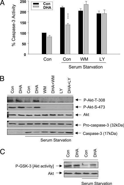
Phosphatidylinositol 3-kinase [PI (3)K]/Akt signaling is a critical pathway in cell survival. Here, we demonstrate a mechanism where membrane alteration by the n-3 fatty acid status affects Akt signaling, impacting neuronal survival. Docosahexaenoic acid (DHA), an n-3 polyunsaturated fatty acid highly enriched in neuronal membranes, promotes neuronal survival by facilitating membrane translocation/activation of Akt through its capacity to increase phosphatidylserine (PS), the major acidic phospholipid in cell membranes. The activation of PI (3)K and phosphatidylsinositol triphosphate formation were not affected by DHA, indicating that membrane interaction of Akt is the event responsible for the DHA effect. Docosapentaenoic acid, which replaces DHA during n-3 fatty acid deficiency, was less effective in accumulating PS and translocating Akt and thus less effective in preventing apoptosis. Consistently, in vivo reduction of DHA by dietary depletion of n-3 fatty acids decreased hippocampal PS and increased neuronal susceptibility to apoptosis in cultures. This mechanism may contribute to neurological deficits associated with n-3 fatty acid deficiency and support protective effects of DHA in pathological models such as brain ischemia or Alzheimer's disease.


Figures

Fig. 1.
Fig. 1.
Effect of DHA on PS accumulation and apoptotic cell death. (A and B) Differential effects of DHA and DPA on PS accumulation (A) and caspase-3 activity induced by serum starvation (B). (C-E) PS-dependent inhibition of apoptosis evidenced by TUNEL positive cells (C) and representative micrographs (D), with respect to PS accumulation altered by polyunsaturated fatty acids and serine depletion (E). (F) Effect of pss1 and pss2 gene silencing on DHA-induced PS accumulation. Caspase-3 activity was expressed as the percentage to the basal value from the nonenriched control. Statistical significance was tested against nonenriched (A and F) or nonenriched serum-free control (B and C). ***, P < 0.001; **, P < 0.01; *, P < 0.05; NS, not significant.
Fig. 2.
Fig. 2.
Role of PI (3)/Akt kinase pathway in DHA's antiapototic effect. (A) The presence of wortmanin (WM) (10 nM) or LY 294002 (LY) (50 μM) during serum starvation abolished DHA's inhibition of caspase-3 activity in serum-starved cells. Results are percentages of the nonenriched control value, and statistical significance was tested against serum-free nonenriched control. ***, P < 0.001. (B) Western blot data showing the reduction of Akt phosphorylation upon serum starvation and the prevention of this reduction by DHA and abolition of DHA's protection by WM and LY. (C) Akt activity maintained by DHA during serum deprivation. Results are representative of at least two independent experiments.
Fig. 3.
Fig. 3.
Effect of DHA and mutation on Akt translocation and phosphorylation. (A) Representative micrographs showing translocation of GFP-AktPH at indicated time points after IGF-1 stimulation with 10 ng/ml in cells supplemented with fatty acids with or without serine. Insets show the fluorescence intensity profile across a transverse section in one cell. (B) Time course of translocation by using the averaged relative R values normalized to the plateau R value for each group (see Materials and Methods). (C) Representative micrographs of IGF-induced translocation of GFP-AktPH mutants (R67A, R69A, R15A, and K20A). (D) Time course of Akt phosphorylation detected with antibodies specific to phospho-Akt Thr-308. The representative data from at least four independent experiments, performed in duplicate, are shown.
Fig. 4.
Fig. 4.
Effect of DHA on the time course of PI (3)K activation or PIP3 formation. (A) In vitro PI phophorylation by immunoprecipitated PI (3)K from control and DHA-enriched cells stimulated with IGF (10 ng/ml) for indicated time periods. (B) PI phosphorylation in control and DHA-enriched cells that were metabolically prelabeled with H332PO4 for 1 h and stimulated for indicated time periods. In both experiments, total lipids were extracted, separated on TLC plates, and exposed to x-ray film. Representative radiographs from two independent experiments are shown.
Fig. 5.
Fig. 5.
Effect of in vivo depletion of n-3 fatty acids on apoptotic cell death and the fatty acid and PS profile in embryonic day 18 hippocampal cultures. (A) Reciprocal replacement of DHA with DPA and significant decrease in PS content by n-3 fatty acid depletion. (B and C) TUNEL-positive cells increased significantly in n-3 fatty acid-deficient culture in basal and overnight trophic factor withdrawal (TFW) conditions (B) as shown in representative micrographs (C). Results represent three independent experiments performed in quadruplicate with ≈1,500 cells evaluated per treatment. The statistical significance was tested against adequate (A) and adequate basal (B) values. *, P < 0.05; ***, P < 0.001.
Fig. 6.
Fig. 6.
Positive effect of DHA on neuronal survival: a schematic model. DHA is provided from astroglia, incorporated into neurons, and promotes PS accumulation by serving as the preferred substrate for PS biosynthesis. Membrane concentration of PS facilitates Akt translocation through interaction with basic residues in the PH domain, resulting in efficient phosphorylation and activation of Akt and suppressing caspase-3 activation and cell death, especially under adverse conditions where PIP3 generation is limited. DPA, replacing DHA in n-3 fatty acid deficiency, is not as effective as DHA in promoting PS accumulation and Akt translocation and, consequently, less effective in supporting cell survival.

References

    1. Dudek, H., Datta, S. R., Franke, T. F., Birnbaum, M. J., Yaou, R., Cooper, G. M., Segal, R. A., Kaplan, D. R. & Greenberg, M. E. (1997) Science 275, 661-665. - PubMed
    1. Crowder, J. & Freeman, R. S. (1998) J. Neurosci. 18, 2933-2943. - PMC - PubMed
    1. Philpott, K. L., McCarthy, M. J., Klippel, A. & Rubin, L. L. (1997) J. Cell Biol. 139, 809-815. - PMC - PubMed
    1. Segal, R. A. (2003) Annu. Rev. Neurosci. 26, 299-330. - PubMed
    1. Salem, N., Jr., Kim, H.-Y. & Yergey, J. A. (1986) in Health Effects of Polyunsaturated Fatty Acids in Seafoods, eds. Simopoulos, A. P. & Kifer, R. R. (Academic, New York), pp. 263-317.

MeSH terms