

Determination of multiple sclerosis plaque size with diffusion-tensor MR Imaging: comparison study with healthy volunteers
- PMID: 16040917
- PMCID: PMC1805677
- DOI: 10.1148/radiol.2362040014
Determination of multiple sclerosis plaque size with diffusion-tensor MR Imaging: comparison study with healthy volunteers
Abstract
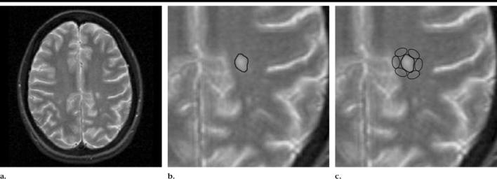
Purpose: To use diffusion-tensor magnetic resonance (MR) imaging to measure involvement of normal-appearing white matter (WM) immediately adjacent to multiple sclerosis (MS) plaques and thus redefine actual plaque size on diffusion-tensor images through comparison with T2-weighted images of equivalent areas in healthy volunteers.
Materials and methods: Informed consent was not required given the retrospective nature of the study on an anonymized database. The study complied with requirements of the Health Insurance Portability and Accountability Act. Twelve patients with MS (four men, eight women; mean age, 35 years) and 14 healthy volunteers (six men, eight women; mean age, 25 years) were studied. The authors obtained fractional anisotropy (FA) values in MS plaques and in the adjacent normal-appearing WM in patients with MS and in equivalent areas in healthy volunteers. They placed regions of interest (ROIs) around the periphery of plaques and defined the total ROIs (ie, plaques plus peripheral ROIs) as abnormal if their mean FA values were at least 2 standard deviations below those of equivalent ROIs within equivalent regions in healthy volunteers. The combined area of the plaque and the peripheral ROI was compared with the area of the plaque seen on T2-weighted MR images by means of a Student paired t test (P = .05).
Results: The mean plaque size on T2-weighted images was 72 mm2 +/- 21 (standard deviation). The mean plaque FA value was 0.285 +/- 0.088 (0.447 +/- 0.069 in healthy volunteers [P < .001]; mean percentage reduction in FA in MS plaques, 37%). The mean plaque size on FA maps was 91 mm2 +/- 35, a mean increase of 127% compared with the size of the original plaque on T2-weighted images (P = .03).
Conclusion: A significant increase in plaque size was seen when normal-appearing WM was interrogated with diffusion-tensor MR imaging. This imaging technique may represent a more sensitive method of assessing disease burden and may have a future role in determining disease burden and activity.
Figures
Similar articles
-
Redefinition of multiple sclerosis plaque size using diffusion tensor MRI.AJR Am J Roentgenol. 2004 Aug;183(2):497-503. doi: 10.2214/ajr.183.2.1830497. AJR Am J Roentgenol. 2004. PMID: 15269047
-
Peritumoral brain regions in gliomas and meningiomas: investigation with isotropic diffusion-weighted MR imaging and diffusion-tensor MR imaging.Radiology. 2004 Aug;232(2):451-60. doi: 10.1148/radiol.2322030959. Epub 2004 Jun 23. Radiology. 2004. PMID: 15215555
-
Multiple sclerosis: diffusion tensor MR imaging for evaluation of normal-appearing white matter.Radiology. 2002 Mar;222(3):729-36. doi: 10.1148/radiol.2223010311. Radiology. 2002. PMID: 11867792
-
Diffusion-weighted MR of the brain: methodology and clinical application.Radiol Med. 2005 Mar;109(3):155-97. Radiol Med. 2005. PMID: 15775887 Review. English, Italian.
-
Diffusion magnetic resonance imaging in multiple sclerosis.J Neurol Neurosurg Psychiatry. 1998 May;64 Suppl 1:S80-4. J Neurol Neurosurg Psychiatry. 1998. PMID: 9647291 Review.
Cited by
-
Diffusion tensor MR imaging of the cervical spinal cord in patients with multiple sclerosis.Eur Radiol. 2007 Oct;17(10):2499-504. doi: 10.1007/s00330-007-0672-4. Epub 2007 May 16. Eur Radiol. 2007. PMID: 17505830
-
Magnetic resonance diffusion tensor imaging for occult lesion detection in multiple sclerosis.Exp Ther Med. 2017 Jan;13(1):91-96. doi: 10.3892/etm.2016.3950. Epub 2016 Dec 2. Exp Ther Med. 2017. PMID: 28123474 Free PMC article.
-
The effect of intravenous gadolinium-DTPA on diffusion-weighted imaging.Neuroradiology. 2006 Jul;48(7):465-70. doi: 10.1007/s00234-006-0091-2. Epub 2006 May 4. Neuroradiology. 2006. PMID: 16673073
-
Cerebral white matter integrity and cognitive aging: contributions from diffusion tensor imaging.Neuropsychol Rev. 2009 Dec;19(4):415-35. doi: 10.1007/s11065-009-9113-2. Epub 2009 Aug 25. Neuropsychol Rev. 2009. PMID: 19705281 Free PMC article. Review.
-
Volumetric assessment of tumor infiltration of adjacent white matter based on anatomic MRI and diffusion tensor tractography.Acad Radiol. 2007 Apr;14(4):431-6. doi: 10.1016/j.acra.2007.01.033. Acad Radiol. 2007. PMID: 17368212 Free PMC article.
References
-
- Miller DH, Grossman RI, Reingold SC, McFarland HF. The role of magnetic resonance techniques in understanding and managing multiple sclerosis. Brain. 1998;121:3–24. - PubMed
-
- McDonald WI, Miller DH, Barnes D. The pathological evolution of multiple sclerosis. Neuropathol Appl Neurobiol. 1992;18:319–334. - PubMed
-
- Stevenson VL, Miller DH, Rovaris M, et al. Primary and transitional progressive MS: a clinical and MRI cross-sectional study. Neurology. 1999;52:839–845. - PubMed
-
- Guo AC, MacFall JR, Provenzale JM. Multiple sclerosis: diffusion tensor MR imaging for evaluation of normal appearing white matter. Radiology. 2002;222:729–736. - PubMed
-
- Werring DJ, Clark CA, Barker GJ, Thompson AJ, Miller DH. Diffusion tensor imaging of lesions and normal-appearing white matter in multiple sclerosis. Neurology. 1999;52:1626–1632. - PubMed
Publication types
MeSH terms
Grants and funding
LinkOut - more resources
Full Text Sources
Medical
