. 2005 Aug;73(8):4512-21.
doi: 10.1128/IAI.73.8.4512-4521.2005.

A toll-like receptor 2-responsive lipid effector pathway protects mammals against skin infections with gram-positive bacteria

Affiliations

A toll-like receptor 2-responsive lipid effector pathway protects mammals against skin infections with gram-positive bacteria

Philippe Georgel et al. Infect Immun. 2005 Aug.

Abstract

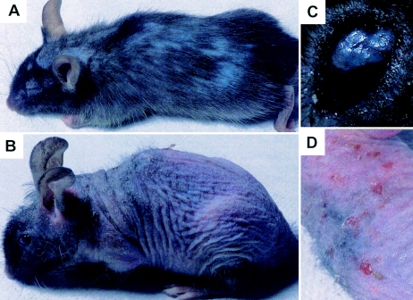
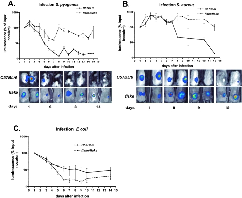
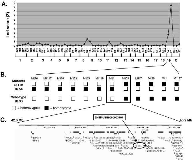
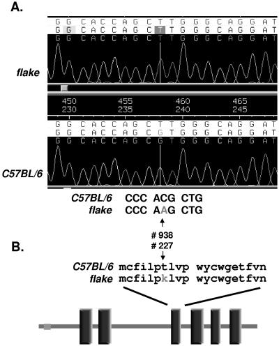
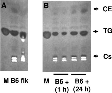
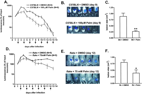
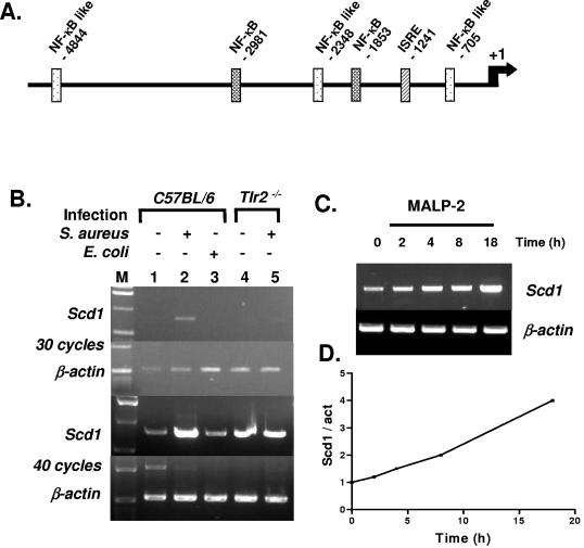
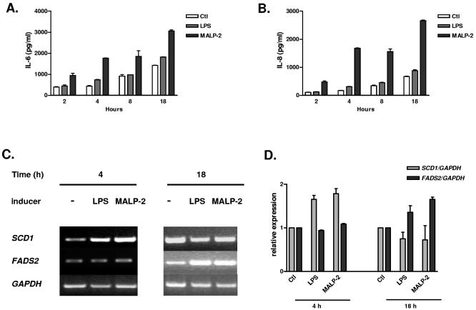
flake (flk), an N-ethyl-N-nitrosourea-induced recessive germ line mutation of C57BL/6 mice, impairs the clearance of skin infections by Streptococcus pyogenes and Staphylococcus aureus, gram-positive pathogens that elicit innate immune responses by activating Toll-like receptor 2 (TLR2). Positional cloning and sequencing revealed that flk is a novel allele of the stearoyl coenzyme A desaturase 1 gene (Scd1). flake homozygotes show reduced sebum production and are unable to synthesize the monounsaturated fatty acids (MUFA) palmitoleate (C(16:1)) and oleate (C(18:1)), both of which are bactericidal against gram-positive (but not gram-negative) organisms in vitro. However, intradermal MUFA administration to S. aureus-infected mice partially rescues the flake phenotype, which indicates that an additional component of the sebum may be required to improve bacterial clearance. In normal mice, transcription of Scd1-a gene with numerous NF-kappaB elements in its promoter--is strongly and specifically induced by TLR2 signaling. Similarly, the SCD1 gene is induced by TLR2 signaling in a human sebocyte cell line. These observations reveal the existence of a regulated, lipid-based antimicrobial effector pathway in mammals and suggest new approaches to the treatment or prevention of infections with gram-positive bacteria.


Figures

FIG. 1.
FIG. 1.
Visible phenotypes observed in flake mutant mice. (A) Six-week-old mouse; (B) 8-month-old mouse; (C) eye infection in an 8-month-old mouse. (D) Magnification of the mouse photograph in panel B highlights severe dermatitis.
FIG. 2.
FIG. 2.
flake mutant mice develop persistent skin infections when exposed to gram-positive bacteria. (A and B) Time course analyses of the bacterial growth in control (C57BL/6) (n = 4) and mutant (flake/flake) (n = 4) animals subcutaneously infected with S. pyogenes (A) or S. aureus (B). (Upper panels) Graphs show results of quantification by luminescence (expressed as a percentage of the luminescence in the initial inoculum) for four animals of each genotype. (Lower panels) Overlay of photographs and light detection for two representative mice for each genotype at the indicated days after inoculation. (C) Infection with E. coli.
FIG. 3.
FIG. 3.
Mapping of the flake mutation. (A) Transgenomic log likelihood ratio [Lod score (Z)] analysis shows a single peak of linkage on mouse chromosome 19. A total of 59 informative markers (horizontal axis) were included in the analysis, and 39 meioses (19 wild-type and 17 mutant animals) were genotyped at all markers. (B) Fine mapping of the distal region of chromosome 19. Analysis of a total of 283 meioses (3 representatives are shown) led to the confinement of the flake mutation between two adjacent markers 2.6 Mb apart. (C) Gene organization at the flake locus according to the Ensembl database. The Scd1 gene is circled.
FIG. 4.
FIG. 4.
Molecular characterization of the flake mutation. (A) Trace file of amplified genomic DNA from homozygous flake mutant mice (top chromatogram) and normal animals (bottom chromatogram). (B) Schematic representation of the SCD1 protein and localization of the flake mutation. Vertical rectangles correspond to transmembrane domains predicted by SMART analysis.
FIG. 5.
FIG. 5.
TLC analysis of the lipid content in wild-type and flake mutant mice. (A) TLC of lipids extracted from skin biopsy specimens of wild-type (B6) or flake (flk) mutant mice. (B) TLC of lipids purified from the skin of wild-type mice (B6 +) 1 h or 24 h after S. aureus subcutaneous infection. Lanes M, markers. Cs, cholesterol; TG, triglycerides; CE, cholesterol esters.
FIG. 6.
FIG. 6.
Palmitoleic acid has antibacterial activity in vivo. (A) Palmitoleate (Palm) injection accelerates bacterial clearance in wild-type mice. Luminescence (expressed as a percentage of the luminescence in the initial inoculum) was measured in control (C57BL/6) mice inoculated with S. aureus (at day 0) and treated by vehicle (DMSO) or palmitoleate injections every 2 days (arrows). (B) Photographs, taken 9 days after S. aureus infection, of control (C57BL/6) mice treated by DMSO (top) or palmitoleate (bottom) injections. (C) Histogram showing the sizes of lesions measured at day 9 after infection in control (B6) mice treated with DMSO or palmitoleate. **, P < 0.01. (D) Palmitoleate treatment of S. aureus-infected flake mice. The protocol is similar to that for panel A, except that we performed 100-μl injections of a 75 mM solution of palmitoleate. (E) Photographs of infected flake mice at day 12 after DMSO (top) or palmitoleate (bottom) treatment. (F) Sizes of lesions (determined at day 12) in infected flk mutants treated with DMSO or palmitoleate. *, P < 0.05.
FIG. 7.
FIG. 7.
Infection- and TLR2-dependent induction of Scd1 gene expression in mice. (A) SignalScan analysis of the Scd1 promoter. NF-κB and ISRE (interferon-stimulated regulatory element) are shown. (B) RT-PCR detection of Scd1 and β-actin transcripts in skin biopsy specimens of uninfected control (C57BL/6) and Tlr2/ animals (lanes 1 and 4) or after infection by S. aureus (lanes 2 and 5) or E. coli (lane 3). PCR products obtained after 30 and 40 cycles are shown. M, size standard. (C) RT-PCR detection of Scd1 and β-actin transcripts in control (0 h) and MALP-induced peritoneal macrophages isolated from wild-type mice after 2, 4, 8, and 18 h. (D) Quantification of the Scd1/β-actin ratio.
FIG. 8.
FIG. 8.
Human sebocytes stimulated with MALP-2 show an inflammatory response and up-regulation of SCD1 and FADS2 genes. (A) IL-6 production is induced in SZ95 cells after MALP-2 treatment (50 ng/ml). LPS stimulation (100 ng/ml) shows minimal effect. (B) Quantification of IL-8 under the same conditions as those in panel A. (C) RT-PCR detection of SCD1 and FADS2 expression 4 and 18 h after LPS and MALP-2 stimulation. GAPDH expression was used as a control. (D) Quantification of the SCD1 and FADS2 signals measured in two independent experiments (± standard errors of the means) after normalization with the GAPDH signal.

References

    1. Alfadley, A., H. K. Al, and A. K. Al. 2003. Ichthyosis follicularis: a case report and review of the literature. Pediatr. Dermatol. 20:48-51. - PubMed
    1. Bunce, C., L. Wheeler, G. Reed, J. Musser, and N. Barg. 1992. Murine model of cutaneous infection with gram-positive cocci. Infect. Immun. 60:2636-2640. - PMC - PubMed
    1. Cohen, P., M. Miyazaki, N. D. Socci, A. Hagge-Greenberg, W. Liedtke, A. A. Soukas, R. Sharma, L. C. Hudgins, J. M. Ntambi, and J. M. Friedman. 2002. Role for stearoyl-CoA desaturase-1 in leptin-mediated weight loss. Science 297:240-243. - PubMed
    1. Egger, S. F., V. Huber-Spitzy, K. Bohler, and C. Scholda. 1995. Isotretinoin administration in treatment of acne vulgaris. A prospective study of the kind and extent of ocular complications. Ophthalmologe 92:17-20. (In German.) - PubMed
    1. Guay, D. R. 2003. Treatment of bacterial skin and skin structure infections. Expert Opin. Pharmacother. 4:1259-1275. - PubMed

Publication types

MeSH terms