. 2005 Aug;73(8):4522-9.
doi: 10.1128/IAI.73.8.4522-4529.2005.

Resistance to intestinal Entamoeba histolytica infection is conferred by innate immunity and Gr-1+ cells

Affiliations

Resistance to intestinal Entamoeba histolytica infection is conferred by innate immunity and Gr-1+ cells

Amon Asgharpour et al. Infect Immun. 2005 Aug.

Abstract

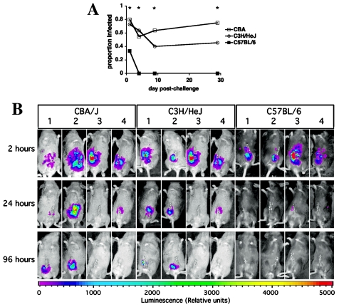
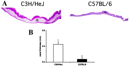
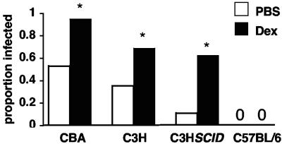
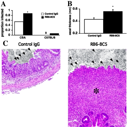
Establishment of intestinal infection with Entamoeba histolytica depends on the mouse strain; C57BL/6 mice are highly resistant, and C3H/HeJ mice are relatively susceptible. We found that resistance to intestinal infection was independent of lymphocyte activity or H-2 haplotype and occurred in the first hours to days postchallenge according to in vivo imaging. At 18 h postchallenge, the ceca of resistant C57BL/6 mice were histologically unremarkable, in contrast to the severe inflammation observed in susceptible C3H/HeJ mice. Comparison of cecal gene expression in C3H/HeJ and C57BL/6 mice demonstrated that there was parasite-induced upregulation of proinflammatory and neutrophil chemotaxis transcripts and there was downregulation of transforming growth factor beta signaling molecules. Pretreatment with dexamethasone abrogated the partial resistance of C3H/HeJ or CBA mice through an innate, lymphocyte-independent mechanism, but it had no effect on the high-level resistance of C57BL/6 mice. Similarly, administration of a neutrophil-depleting anti-Gr-1 monoclonal antibody (RB6-8C5) decreased the partial resistance of CBA mice and led to severe pathology compared to control antibody-treated mice, but it had no effect on C57BL/6 resistance. These data indicate that there are discrete mechanisms of innate resistance to E. histolytica depending on the host background and, in contrast to other reports, imply that neutrophils are protective and not damaging in intestinal amebiasis.


Figures

FIG. 1.
FIG. 1.
Resistance to E. histolytica occurs within hours in the mouse intestine. (A) CBA, C3H/HeJ, and C57BL/6 mice were challenged on day 0 and sacrificed sequentially on days 1, 4, 9, and 29. The proportions of mice infected as confirmed by culture and histopathology are shown (n = 10 to n = 12 for each mouse strain on each day). An asterisk indicates that the P value is <0.04 for a comparison of the rates of infection of CBA or C3H/HeJ mice and C57BL/6 mice; the infection rates were not significantly different for CBA or C3H/HeJ mice at any time. (B) CBA, C3H/HeJ, and C57BL/6 mice were intracecally inoculated with luciferase-expressing E. histolytica trophozoites and serially imaged with a charge-coupled device camera at 2, 24, and 96 h after intraperitoneal administration of luciferin. Mice were sacrificed at 96 h for confirmation of infection. Four representative mice of each strain are shown. CBA mice 1 and 2 and C3H mice 1 and 2 were infected based on histology and culture, while all other mice were uninfected. Infection was limited to the cecum. Luciferase-expressing trophozoites exhibited no luminescence without administration of luciferin, and trophozoites transfected with the control plasmid did not luminesce (data not shown).
FIG. 2.
FIG. 2.
C3H/HeJ mice exhibit rapid intestinal inflammation in response to acute E. histolytica challenge. (A) Histology 18 h after intracecal challenge with E. histolytica trophozoites showed diffuse cecal inflammation with submucosal edema (asterisk) in the susceptible C3H/HeJ strain compared with the normal histology in the C57BL/6 mouse (hematoxylin and eosin staining). Magnification, ×20. (B) Cross-sectional cecal thickness was measured in both strains at 18 h. The data are means and standard errors (n = 8). An asterisk indicates that the P value is 0.007.
FIG. 3.
FIG. 3.
Dexamethasone decreases innate resistance to intestinal E. histolytica infection in susceptible strains. CBA, C3H/HeJ, C3H SCID, and C57BL/6 mice were treated with 0.2 mg dexamethasone (Dex) or PBS on days −3, −2, −1, and 0 relative to intracecal challenge. The proportions of mice infected upon sacrifice are shown (n = 15, n = 20, n = 34, n = 29, n = 9, n = 8, n = 6, and n = 8 for the groups shown from left to right on the x axis) An asterisk indicates that the P value is ≤0.05 compared with PBS-treated mice. C3H/HeJ mice were sacrificed at 30 days postchallenge, and all other mice were sacrificed at 10 days postchallenge.
FIG. 4.
FIG. 4.
Innate resistance to intestinal E. histolytica infection is diminished by neutrophil-depleting anti-Gr-1 MAb administration in CBA mice. (A) Six- to 10-week-old mice with the CBA and C57BL/6 mice background were given 300 μg of anti-Gr-1 MAb RB6-8C5 or control rat IgG on days −2, −1, and 2 relative to intracecal challenge. Neutrophil depletion was confirmed in the peripheral blood on days 0 and 6 and in the intestine by the MPO assay on day 6. Mice were sacrificed on day 6 for evaluation of infection by histology and culture. An asterisk indicates that the P value is 0.01 for a comparison of anti-Gr-1 MAb-treated and control IgG-treated CBA mice (n = 22, n = 26, n = 13, and n = 13 for the four groups shown from left to right on the x axis). (B) In infected CBA mice, cecal thickness was compared for the anti-Gr-1MAb-treated and control IgG-treated groups. The data are means and standard errors (n = 12 and n = 23 for infected, control IgG-treated mice and infected, anti-Gr-1 MAb-treated mice, respectively) An asterisk indicates that the P value is ≤0.01. (C) Representative photomicrographs showing that there was significant destruction of mucosal architecture with infiltration (asterisk) in a neutrophil-depleted animal. Luminal contents are black and white, and trophozoites are indicated by arrows.

References

    1. Ashburner, M., C. A. Ball, J. A. Blake, D. Botstein, H. Butler, J. M. Cherry, A. P. Davis, K. Dolinski, S. S. Dwight, J. T. Eppig, M. A. Harris, D. P. Hill, L. Issel-Tarver, A. Kasarskis, S. Lewis, J. C. Matese, J. E. Richardson, M. Ringwald, G. M. Rubin, and G. Sherlock. 2000. Gene ontology: tool for the unification of biology. The Gene Ontology Consortium. Nat. Genet. 25:25-29. - PMC - PubMed
    1. Auphan, N., J. A. DiDonato, C. Rosette, A. Helmberg, and M. Karin. 1995. Immunosuppression by glucocorticoids: inhibition of NF-kappa B activity through induction of I kappa B synthesis. Science 270:286-290. - PubMed
    1. Blessmann, J., I. K. Ali, P. A. Nu, B. T. Dinh, T. Q. Viet, A. L. Van, C. G. Clark, and E. Tannich. 2003. Longitudinal study of intestinal Entamoeba histolytica infections in asymptomatic adult carriers. J. Clin. Microbiol. 41:4745-4750. - PMC - PubMed
    1. Bliss, S. K., L. C. Gavrilescu, A. Alcaraz, and E. Y. Denkers. 2001. Neutrophil depletion during Toxoplasma gondii infection leads to impaired immunity and lethal systemic pathology. Infect. Immun. 69:4898-4905. - PMC - PubMed
    1. Burchard, G. D., G. Prange, and D. Mirelman. 1993. Interaction between trophozoites of Entamoeba histolytica and the human intestinal cell line HT-29 in the presence or absence of leukocytes. Parasitol. Res. 79:140-145. - PubMed

Publication types