. 2005 Aug;73(8):4530-8.
doi: 10.1128/IAI.73.8.4530-4538.2005.

Modulation of the lung inflammatory response to serotype 8 pneumococcal infection by a human immunoglobulin m monoclonal antibody to serotype 8 capsular polysaccharide

Affiliations

Modulation of the lung inflammatory response to serotype 8 pneumococcal infection by a human immunoglobulin m monoclonal antibody to serotype 8 capsular polysaccharide

Tamika Burns et al. Infect Immun. 2005 Aug.

Abstract

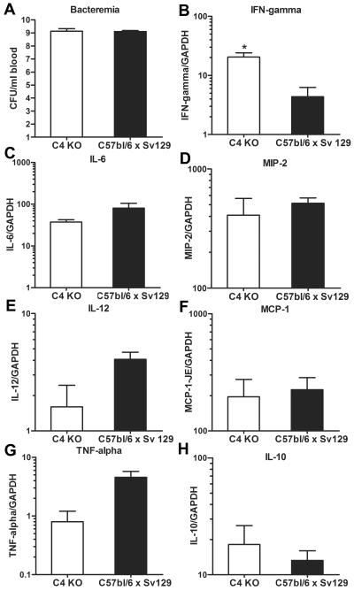
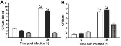
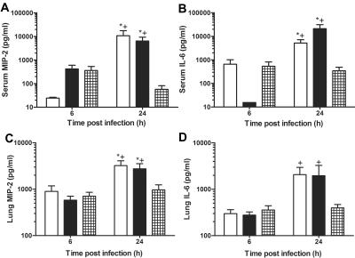
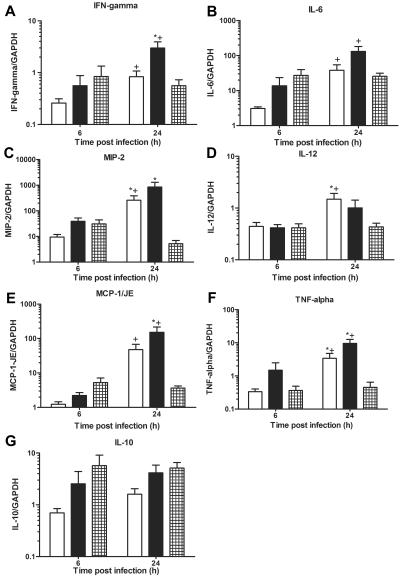
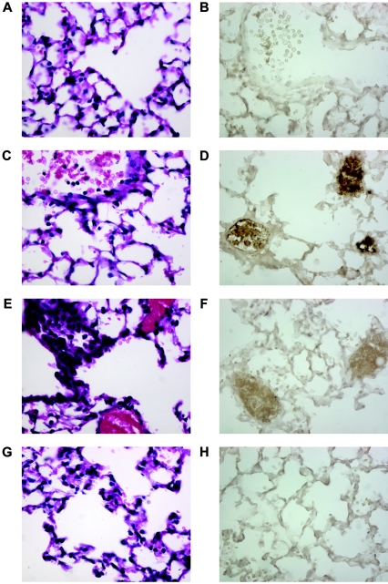
The human monoclonal antibody to serotype 8 pneumococcal capsular polysaccharide D11 [immunoglobulin M(kappa)] protects wild-type and complement component 4 knockout (C4 KO) mice against lethal intratracheal challenge with serotype 8 pneumococcus, but it does not promote polymorphonuclear leukocyte (PMN)-mediated pneumococcal killing in vitro. In this study, we investigated the effect of D11 on the blood and lung bacterial burdens and the serum and lung expression of inflammatory chemokines and cytokines in an intratracheal challenge model with serotype 8 pneumococcus in C4 KO mice. Pneumococcus was not detected in the blood of D11-treated mice, whereas control mice had high-grade bacteremia with >10(7) CFU. Control mice had a >5-log increase in lung CFU and D11-treated mice manifested a nearly 3-log increase in lung CFU compared to the original inoculum 24 h after infection. Serum and lung levels of soluble macrophage inflammatory protein 2 (MIP-2) and interleulin-6 (IL-6) as measured by an enzyme-linked immunosorbent assay were lower in D11-treated mice than in control mice 24 h after infection. Real-time PCR was performed to examine lung mRNA chemokine and cytokine expression. The results showed that D11-treated mice had significantly less gamma interferon, MIP-2, IL-12, monocyte chemoattractant protein 1/JE, and tumor necrosis factor alpha expression than control mice 24 h after infection. Histopathology and immunohistochemical staining of lung tissues revealed that D11-treated mice had less inflammation, fewer PMNs, and less myeloperoxidase staining than control mice 24 h after infection. These findings suggest that the efficacy of certain serotype-specific antibodies against pneumococcal pneumonia could be associated with modulation of the lung inflammatory response and a reduction in host damage.


Figures

FIG. 1.
FIG. 1.
Bacteremia and cytokine and chemokine expression in background strain and C4 KO mice: cytokine lung expression and number of CFU of type 8 pneumococcus in whole blood of C4 KO (open bars) and C57BL/6 × Sv129 (solid bars) mice infected i.t. with either 20 or 100 CFU of serotype 8 pneumococcus. (A) Bacteremia. (B) IFN-γ. (C) IL-6. (D) MIP-2. (E) IL-12. (F) MCP-1/JE. (G) TNF-α. (H) IL-10. The bars indicate the means, and the errors bars indicate the standard errors of the means. An asterisk indicates that the P value is 0.02, as determined by an unpaired t test (n = 3). GAPDH, glyceraldehyde-3-phosphate dehydrogenase.
FIG. 2.
FIG. 2.
Bacteremia and lung CFU in C4 KO mice infected i.t. with serotype 8 pneumococcus: CFU in blood (A) and lungs (B) obtained from C4 KO mice inoculated with serotype 8 pneumococcus and PBS (open bars), 1 μg of IgM (solid bars), or 1 μg of D11 (cross-hatched bars) at 6 and 24 h after infection. An asterisk indicates that the P value is <0.05 for a comparison of control mice at 24 h to D11-treated mice, as determined by an unpaired t test; a plus sign indicates that the P value is < 0.05 for a comparison of the numbers of CFU at 6 h and 24 h after infection, as determined by an unpaired t test (n = 12 to 15 mice per time).
FIG. 3.
FIG. 3.
Serum and lung soluble MIP-2 and IL-6 levels in C4 KO mice infected i.t. with serotype 8 pneumococcus: serum MIP-2 and IL-6 (A and B) and lung MIP-2 and IL-6 (C and D) protein levels as determined by ELISA in PBS-treated mice (open bars), IgM-treated mice (solid bars), and D11-treated mice (cross-hatched bars) at 6 and 24 h after infection. The bars indicate the means of three independent experiments, and the error bars indicate the standard errors of the means. An asterisk indicates that the P value is <0.05 for a comparison of the control mice to D11-treated mice at 24 h after infection, as determined by an unpaired t test; a plus sign indicates that the P value is <0.05 for a comparison of the levels at 6 h and 24 h after infection, as determined by an unpaired t test (n = 12 to 15 mice per time).
FIG. 4.
FIG. 4.
Lung mRNA chemokine and cytokine expression in C4 KO mice infected i.t. with serotype 8 pneumococcus: lung mRNA chemokine and cytokine expression as determined by real-time PCR for mice inoculated i.t. with serotype 8 pneumococcus and PBS-treated mice (open bars), IgM-treated mice (solid bars), or D11-treated mice (cross-hatched bars) at 6 and 24 h after infection. (A) IFN-γ. (B) IL-6. (C) MIP-2. (D) IL-12. (E) MCP-1/JE. (F) TNF-α. (G) IL-10. The bars indicate the means of four independent experiments, and the error bars indicate the standard errors of the means. The y axis shows the mRNA concentrations of the mediators normalized to the concentration of glyceraldehyde-3-phosphate dehydrogenase (GAPDH). An asterisk indicates that the P value is <0.05 for a comparison of control and D11-treated mice at 24 h after infection, as determined by an unpaired t test; a plus sign indicates that the P value is <0.05 for a comparison of the mRNA levels at 6 h and 24 h after infection, as determined by an unpaired t test (n = 10 to 12 mice per time).
FIG. 5.
FIG. 5.
Histopathological appearance of C4 KO mice infected i.t. with type 8 pneumococcus: fixed tissue sections obtained 6 and 24 h after infection and stained with H&E and anti-myeloperoxidase antibody. (A) IgM, 6 h, H&E. (B) IgM, 6 h, MPO. (C) D11, 6 h, H&E. (D) D11, 6 h, MPO. (E) IgM, 24 h, H&E. (F) IgM, 24 h, MPO. (G) D11, 24 h, H&E. (H) D11, 24 h, MPO. Magnification, ×40. The images are representative of two sections per time.

Similar articles

Cited by

References

    1. Akilesh, S., D. J. Shaffer, and D. Roopenian. 2003. Customized molecular phenotyping by quantitative gene expression and pattern recognition analysis. Genome Res. 13:1719-1727. - PMC - PubMed
    1. Ali, F., M. E. Lee, F. Iannelli, G. Pozzi, T. J. Mitchell, R. C. Read, and D. H. Dockrell. 2003. Streptococcus pneumoniae-associated human macrophage apoptosis after bacterial internalization via complement and Fcgamma receptors correlates with intracellular bacterial load. J. Infect. Dis. 188:1119-1131. - PubMed
    1. Anttila, M., M. Voutilainen, V. Jantti, J. Eskola, and H. Kayhty. 1999. Contribution of serotype-specific IgG concentration, IgG subclasses and relative antibody avidity to opsonophagocytic activity against Streptococcus pneumoniae. Clin. Exp. Immunol. 118:402-407. - PMC - PubMed
    1. Arva, E., and B. Andersson. 1999. Induction of phagocyte-stimulating and Th1-promoting cytokines by in vitro stimulation of human peripheral blood mononuclear cells with Streptococcus pneumoniae. Scan. J. Immunol. 49:417-423. - PubMed
    1. Austrian, R., R. M. Douglas, G. Schiffman, A. M. Coetzee, H. J. Koornhof, S. Hayden-Smith, and R. D. Reid. 1976. Prevention of pneumococcal pneumonia by vaccination. Trans. Assoc. Am. Physicians 89:184-194. - PubMed

Publication types

MeSH terms