. 2005 Aug;73(8):4571-80.
doi: 10.1128/IAI.73.8.4571-4580.2005.

Virulence of the fungal pathogen Candida albicans requires the five isoforms of protein mannosyltransferases

Affiliations

Virulence of the fungal pathogen Candida albicans requires the five isoforms of protein mannosyltransferases

Mahmoud Rouabhia et al. Infect Immun. 2005 Aug.

Abstract

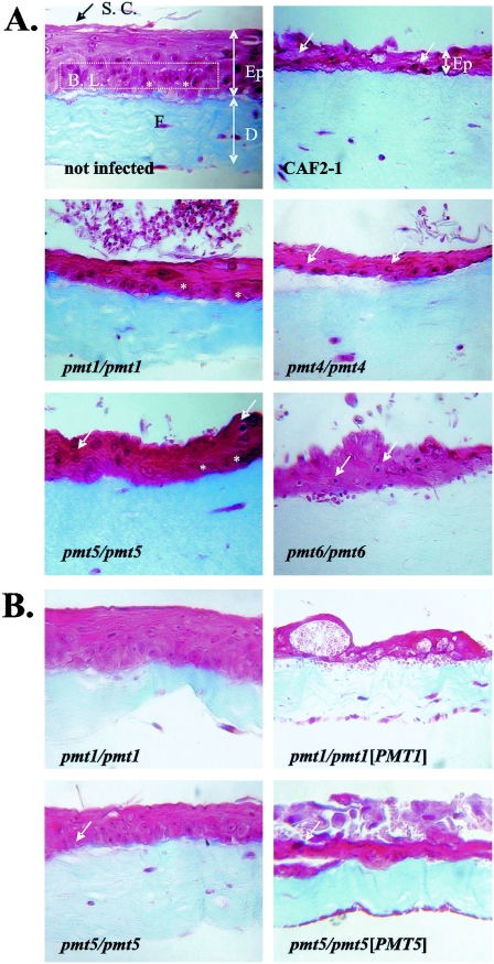
The PMT gene family in Candida albicans encodes five isoforms of protein mannosyltransferases (Pmt proteins Pmt1p, Pmt2p, Pmt4p, Pmt5p, and Pmt6p) that initiate O mannosylation of secretory proteins. We compared virulence characteristics of pmt mutants in two complex, three-dimensional models of localized candidiasis, using reconstituted human epithelium (RHE) and engineered human oral mucosa (EHOM); in addition, mutants were tested in a mouse model of hematogenously disseminated candidiasis (HDC). All pmt mutants showed attenuated virulence in the HDC model and at least one model of localized candidiasis. The pmt5 mutant, which lacks in vitro growth phenotypes, was less virulent in the EHOM and HDC assays but had no consistent phenotype in the RHE assay. In contrast, the pmt4 and pmt6 mutants were less virulent in the RHE and HDC assays but not in the EHOM assay. The results stress the contribution of all Pmt isoforms to the virulence of C. albicans and suggest that the importance of individual Pmt isoforms may differ in specific host niches. We propose that Pmt proteins may be suitable targets for future novel classes of antifungal agents.


Figures

FIG. 1.
FIG. 1.
Infection of RHE by C. albicans. A total of 2 × 10⁶ C. albicans cells were used to infect the RHE, which was examined at 24 h by light microscopy. The strains used for infection were CAI4 (PMT/PMT), CAP1-3121(pRC18) (pmt1/pmt1), CAP1-3121(pCT30) (pmt1/pmt1[PMT1]), CAP4-2164(pRC18) (pmt4/pmt4), CAP4-2164(pSP1) (pmt4/pmt4[PMT4]), CAP2-2391(pRC18) (pmt6/pmt6), and CAP2-2391(pCT34) (pmt6/pmt6[PMT6]). Note that epithelial invasion caused by C. albicans CAI4 and PMT-reconstituted strains (dark spheres and filaments) is accompanied by vacuolization and edema of all keratinocyte layers.
FIG.2.
FIG.2.
Histological appearance of EHOM not infected or infected with C. albicans strains. An uninfected EHOM has an epidermis (Ep) containing an outmost stratum corneum layer (S.C.) and a basal layer (B. L.), as well as a collagen-containing dermis (D) that contains some fibroblasts (F). Cuboidal cells in the basal layer are indicated by asterisks, and large differentiated cells are indicated by white arrows. Following 22 h of infection, biopsies were taken from each EHOM, and Masson trichrome staining was performed. Sections of stained tissues were observed with an optical microscope and photographed. Representative photographs from three different experiments are shown (magnification, ×250). (A) Comparison of URA3-reconstituted pmt mutants with control strain CAF2-1. Mutants SPCa2 (pmt1), SPCa6 (pmt4), SPCa10 (pmt5), and SPCa8 (pmt6) were used for infection. (B) Comparison of PMT-reconstituted mutants and unreconstituted mutants. Strains CAP1-3121(pRC18) (pmt1/pmt1), CAP1-3121(pCT30) (pmt1/pmt1[PMT1]), P5-5744(pRC18) (pmt5/pmt5), and P5-5744(pBK1) (pmt5/pmt5) were compared.
FIG. 3.
FIG. 3.
Virulence of C. albicans in an HDC model. CD2F1 mice were infected with C. albicans strains (1 × 10⁶ cells/mouse). (A) Survival curves determined by the Kaplan-Meier method. Strains CAF2-1 (•), CAP4-2164(pSP1) (pmt4/pmt4 [PMT4]) (▪), CAP4-2164(pRC18) (pmt4/pmt4) (▴), P5-5744(pBK1) (pmt5/pmt5 [PMT5]) (▾), and P5-5744(pRC18) (pmt5/pmt5) (⧫) were tested. (B) Viable C. albicans cells in the kidneys and brain. Mice were treated with C. albicans strains (1 × 10⁶ cells/mouse). At different times, mice were killed, and viable yeasts (CFU) in the kidneys (grey bars) and brain (black bars) were counted by plating samples of homogenized tissues on Sabouraud agar. The data are the means ± standard deviations for five mice.

Similar articles

Cited by

References

    1. Aoki, S., S. Ito-Kuwa, K. Nakamura, K. Ninomiya, and V. Vidotto. 1994. Extracellular proteolytic activity of Cryptococcus neoformans. Mycopathologia 128:143-150. - PubMed
    1. Buurman, E. T., C. Westwater, B. Hube, A. J. P. Brown, F. C. Odds, and N. A. R. Gow. 1998. Molecular analysis of Ca Mnt1p, a mannosyl transferase important for adhesion and virulence of Candida albicans. Proc. Natl. Acad. Sci. USA 95:7670-7675. - PMC - PubMed
    1. Cameron, B. J., and L. J. Douglas. 1996. Blood group glycolipids as epithelial cell receptors for Candida albicans. Infect. Immun. 64:891-896. - PMC - PubMed
    1. Chaffin, W. L., J. L. Lopez-Ribot, M. Casanova, D. Gozalbo, and J. P. Martinez. 1998. Cell wall and secreted proteins of Candida albicans: identification, function, and expression. Microbiol. Mol. Biol. Rev. 62:130-180. - PMC - PubMed
    1. Cheng, S., M. H. Nguyen, Z. Zhang, H. Jia, M. Handfield, and C. J. Clancy. 2003. Evaluation of the roles of four Candida albicans genes in virulence by using gene disruption strains that express URA3 from the native locus. Infect. Immun. 71:6101-6103. - PMC - PubMed

Publication types

MeSH terms

LinkOut - more resources