. 2005 Aug;73(8):4596-606.
doi: 10.1128/IAI.73.8.4596-4606.2005.

Heparin stimulates Staphylococcus aureus biofilm formation

Affiliations

Heparin stimulates Staphylococcus aureus biofilm formation

Robert M Q Shanks et al. Infect Immun. 2005 Aug.

Abstract

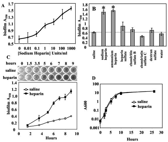
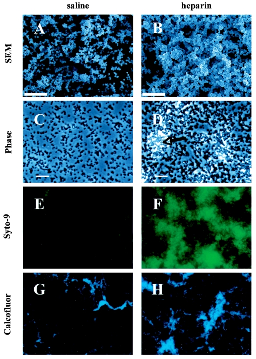
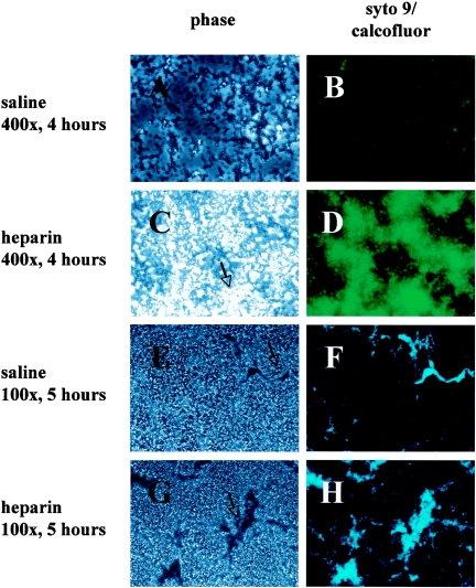
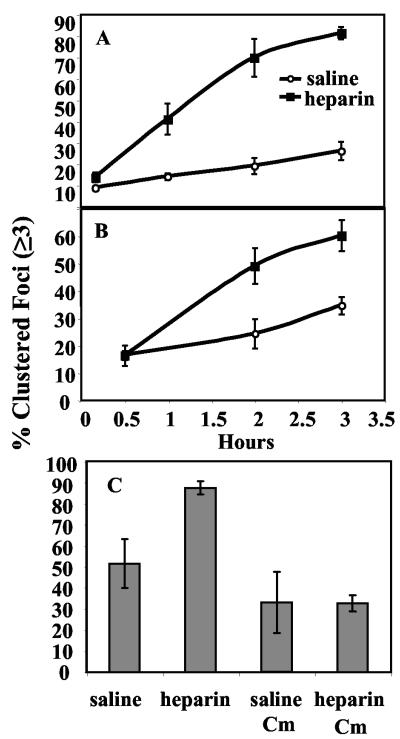
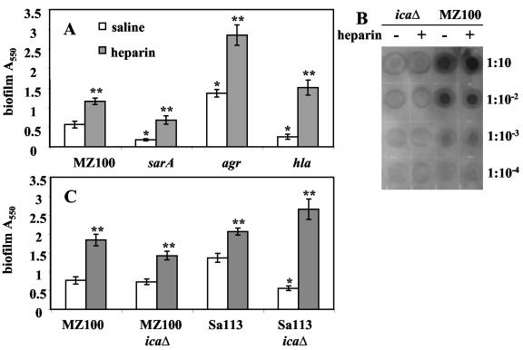
Heparin, known for its anticoagulant activity, is commonly used in catheter locks. Staphylococcus aureus, a versatile human and animal pathogen, is commonly associated with catheter-related bloodstream infections and has evolved a number of mechanisms through which it adheres to biotic and abiotic surfaces. We demonstrate that heparin increased biofilm formation by several S. aureus strains. Surface coverage and the kinetics of biofilm formation were stimulated, but primary attachment to the surface was not affected. Heparin increased S. aureus cell-cell interactions in a protein synthesis-dependent manner. The addition of heparin rescued biofilm formation of hla, ica, and sarA mutants. Our data further suggest that heparin stimulation of biofilm formation occurs neither through an increase in sigB activity nor through an increase in polysaccharide intracellular adhesin levels. These finding suggests that heparin stimulates S. aureus biofilm formation via a novel pathway.


Figures

FIG. 1.
FIG. 1.
Sodium heparin increases the adherence of S. aureus to polystyrene. A. Dose response. Serial dilutions of sodium heparin were added to cultures and biofilms were allowed to form for 16 h, at which time nonadherent cells were removed by vigorous washing. Adherent cells were stained with crystal violet. Spectrophotometric analysis of solubilized crystal violet is shown as a function of increasing sodium heparin concentration in U/ml. B. Biofilm formation in response to various charged molecules and glycosaminoglycans. Microtiter biofilms were analyzed as above, and significant differences from the saline-alone control are shown with an asterisk (P < 0.01). C. Adherence kinetics. The effect of sodium heparin (1,000 U/ml, 10% vol/vol) or saline (10% vol/vol) upon biofilm formation was assessed over time. A photograph of crystal violet-stained biofilms is shown at the top of the panel and spectrophotometric results are charted with respect to time at the bottom of the panel. D. Growth curve. The effect of heparin (1,000 U/ml) on planktonic growth in TSB plus glucose was analyzed and plotted versus time.
FIG. 2.
FIG. 2.
Sodium heparin enhances S. aureus biofilm formation. The effect of heparin on the formation of S. aureus MZ100 biofilms on abiotic surfaces was assessed microscopically. Scanning electron micrographs of 12-hour-old S. aureus biofilms on polyvinylchloride are shown with (B) and without (A) heparin (12,500× magnification, bar = 10 μm). S. aureus biofilms (4 h) formed on polystyrene were viewed with phase-contrast microscopy (C and D, bar = 20 μm, the arrow indicates a phase-bright microcolony), or were stained with a fluorescent bacterial stain (Styo-9) with a 250-ms exposure and at 400× magnification with epifluorescent microscopy (E, without heparin, and F, with heparin at 1,000 U/ml). The 5-hour-old biofilms were stained with calcofluor and viewed with epifluorescent microscopy at 100× magnification, with (H) and without (G) heparin.
FIG. 3.
FIG. 3.
Sodium heparin enhances S. aureus biofilm formation. The effect of heparin formation of S. aureus (MZ100) biofilms on abiotic surfaces was assessed microscopically. S. aureus biofilms (4 hours) formed on polystyrene were viewed with phase 2 microscopy (A, without heparin, and C, with heparin, the arrow indicates one of several phase-bright microcolonies), or were stained with a fluorescent bacterial stain (Styo-9) with a 250-ms exposure and at 400× magnification with epifluorescent microscopy (B, without heparin, and D, with heparin at 1,000 U/ml). The 5-h-old biofilms were stained with calcofluor and viewed with phase 2 microscopy (E, without heparin, and G, with heparin, the arrow indicates a macrocolony), or epifluorescent microscopy at 100× magnification, without (F) or with (H) heparin.
FIG. 4.
FIG. 4.
Heparin indirectly stimulates S. aureus cell-cell interactions. The effect of heparin on cell-cell interactions was assessed using phase-contrast microscopy with strain MZ100. Foci were counted and classified as either clusters (≥3 cells/focus) or nonclusters (1 or 2 cells per focus). A. Planktonic-phase clustering: aliquots were removed at the times indicated, viewed microscopically, assessed for cell-cell interactions, and plotted as percent of foci in clusters as a function of time (n = 1,492 foci). B. Surface clustering: cells attached to a polystyrene surface were microscopically assayed (n = 18,632 foci). The percent of foci in clusters are plotted as a function of time. C. Clustering without protein synthesis: a planktonic culture was treated as above except that chloramphenicol (30 μg/ml) was added to inhibit protein synthesis. A double inoculum of cells was added at the onset of the experiment to cultures exposed to chloramphenicol to control for the difference in cell number that could affect the frequency of cell-cell interactions. At 180 min, samples from four wells per category were assessed microscopically for cell-cell interactions (n = 2,025 foci).
FIG. 5.
FIG. 5.
Effect of heparin on known biofilm formation mutants. A. Mutants (gene names noted) were assessed for biofilm formation in the microtiter dish assay (8-h biofilms) with heparin at 1,000 U/ml (shaded bars) or with saline (white bars). B. Relative PIA levels were determined in response to heparin at 1,000 U/ml. Tenfold serial dilutions of protease-treated whole-cell lysates were immunoblotted with anti-PIA antibodies. C. Isogenic pairs of wild-type and ica mutants in the MZ100 and Sa113 backgrounds were assessed for biofilm formation in the microtiter dish assay (9-h biofilms). One asterisk signifies statistical significance (P < 0.05) between a wild-type strain and its isogenic mutant strain without heparin; two asterisks signify statistical significance (P < 0.05) between strains with and without heparin.

Similar articles

Cited by

References

    1. Arnow, P. M., E. M. Quimosing, and M. Beach. 1993. Consequences of intravascular catheter sepsis. Clin. Infect. Dis. 16:778-784. - PubMed
    1. Bateman, B. T., N. P. Donegan, T. M. Jarry, M. Palma, and A. L. Cheung. 2001. Evaluation of a tetracycline-inducible promoter in Staphylococcus aureus in vitro and in vivo and its application in demonstrating the role of sigB in microcolony formation. Infect. Immun. 69:7851-7857. - PMC - PubMed
    1. Beenken, K. E., J. S. Blevins, and M. S. Smeltzer. 2003. Mutation of sarA in Staphylococcus aureus limits biofilm formation. Infect. Immun. 71:4206-4211. - PMC - PubMed
    1. Beenken, K. E., P. M. Dunman, F. McAleese, D. Macapagal, E. Murphy, S. J. Projan, J. S. Blevins, and M. S. Smeltzer. 2004. Global gene expression in Staphylococcus aureus biofilms. J. Bacteriol. 186:4665-4684. - PMC - PubMed
    1. Bertani, G. 1951. Studies on lysogenesis. I. The mode of phage liberation by lysogenic Escherichia coli. J. Bacteriol. 62:293-300. - PMC - PubMed

Publication types

MeSH terms