. 2005 Aug;73(8):4653-67.
doi: 10.1128/IAI.73.8.4653-4667.2005.

Illustration of pneumococcal polysaccharide capsule during adherence and invasion of epithelial cells

Affiliations

Illustration of pneumococcal polysaccharide capsule during adherence and invasion of epithelial cells

Sven Hammerschmidt et al. Infect Immun. 2005 Aug.

Abstract

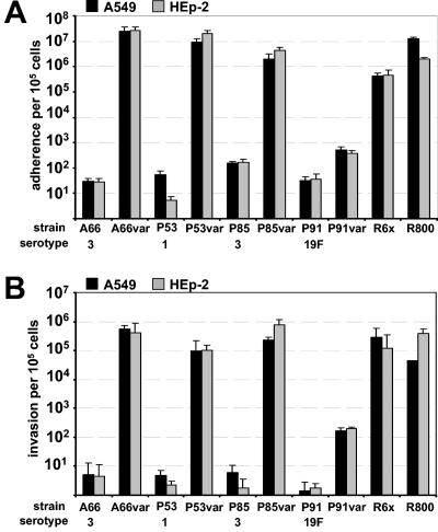
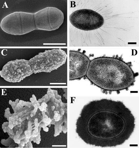
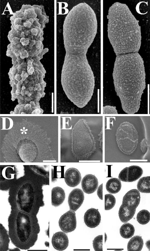
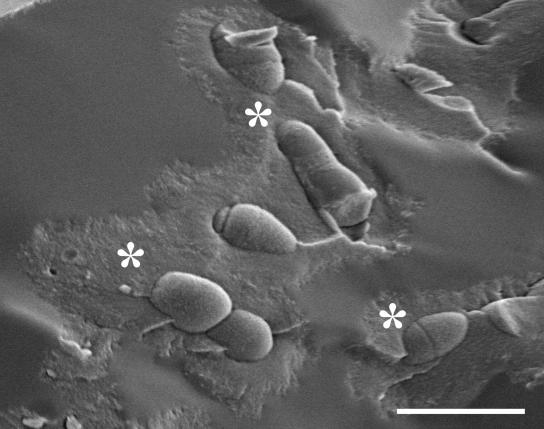
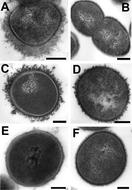
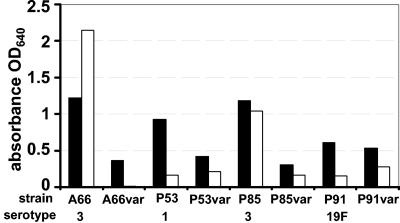
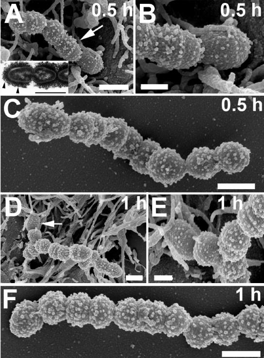
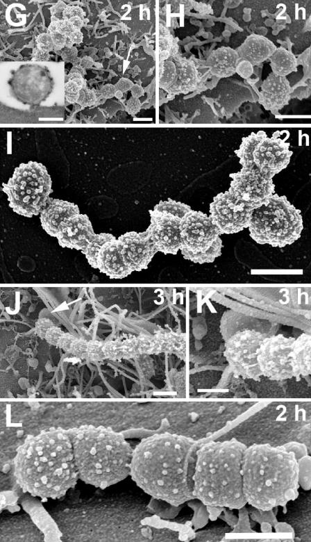
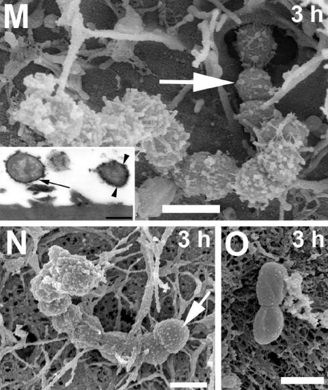
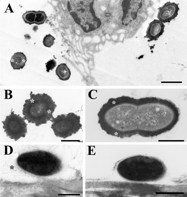
The capsular polysaccharide of Streptococcus pneumoniae represents an important virulence factor and protects against phagocytosis. In this study the amount of capsular polysaccharide present on the bacterial surface during the infection process was illustrated by electron microscopic studies. After infection of A549 cells (type II pneumocytes) and HEp-2 epithelial cells a modified fixation method was used that allowed visualization of the state of capsule expression. This modified fixation procedure did not require the use of capsule-specific antibodies. Visualization of pneumococci in intimate contact and invading cells demonstrated that pneumococci were devoid of capsular polysaccharide. Pneumococci not in contact with the cells did not show alterations in capsular polysaccharide. After infection of the cells, invasive pneumococci of different strains and serotypes were recovered. Single colonies of these recovered pneumococci exhibited an up-to-10(5)-fold-enhanced capacity to adhere and an up-to-10(4)-fold-enhanced capacity to invade epithelial cells. Electron microscopic studies using a lysine-ruthenium red (LRR) fixation procedure or cryo-field emission scanning electron microscopy revealed a reduction in capsular material, as determined in detail for a serotype 3 pneumococcal strain. The amount of polysaccharide in the serotype 3 capsule was also determined after intranasal infection of mice. This study illustrates for the first time the phenotypic variation of the polysaccharide capsule in the initial phase of pneumococcal infections. The modified LRR fixation allowed monitoring of the state of capsule expression of pathogens during the infectious process.


Figures

FIG. 1.
FIG. 1.
Efficiency of adherence and invasion of pneumococcal variants recovered from epithelial cells. Equal amounts of wild-type pneumococci and their variants were used in the cell culture infection assay. Adherence (A) and invasion (B) were measured microscopically by double immunofluorescence. var, recovered pneumococci. The data are the means ± standard errors of five independent experiments.
FIG. 2.
FIG. 2.
Comparison of different fixation methods for visualization of the capsule. Conventional fixation with formaldehyde and glutaraldehyde resulted in a total loss of the capsule of serotype 3 pneumococcus strain A66, as demonstrated by FESEM (A) and in ultrathin sections (B). Addition of ruthenium red in the fixation protocol resulted in some remaining structural material of the capsule on the pneumococcal surface (C and D). A well-preserved capsular structure was observed when a lysine-acetate-based ruthenium red-osmium fixation protocol was used (E and F). (A, C, and E) Bars = 0.5 μm. (B, D, and F) Bars = 0.1 μm.
FIG. 3.
FIG. 3.
Comparison of capsular structures of serotype 3 strain A66 and recovered pneumococci. Pneumococcal variants of strain A66 recovered from HEp-2 cells (B, E, and H) and A459 cells (C, F, and I) were devoid of any visible capsular material, whereas strain A66 exhibited a dense capsular layer (A, D, and G). These observations were made by using three different methods, conventional FESEM (A to C), cryo-FESEM (D to F), and analysis of ultrathin sections (G to I) after LRR fixation. Bars = 0.5 μm.
FIG. 4.
FIG. 4.
Visualization of the vitrified pneumococcal capsule using cryo-FESEM after LRR fixation. Cryo-FESEM analysis revealed a dense thick capsule (asterisks) around serotype 3 strain A66, which is comparable to the capsule visualized in the ultrathin sections. Bar = 2 μm.
FIG. 5.
FIG. 5.
Capsular structures in different serotypes. LRR fixation allowed detection of capsular structures in serotype 1 (wild-type strain P53) (A) and serotype 19F (wild-type strain P91) (C). Furthermore, the fixation protocol unequivocally demonstrated the absence of capsular material in the isolated intracellular variants of serotype 1 (B), loss of capsular material in variants of serotype 19F (D), and the absence of capsular structures in nonencapsulated strains R6x (E) and R800 (F). Bars = 0.25 μm.
FIG. 6.
FIG. 6.
Quantification of bacterium-associated polysaccharides or polysaccharides in culture supernatants. The differences in bacterium-associated polysaccharides (solid bars) or polysaccharides in the culture supernatants (open bars) were assessed for wild-type pneumococci and pneumococcal variants (var) recovered from epithelial cells. The total amount of acidic polysaccharides (bacterium associated or in the supernatant) was measured by determining the optical density at 640 nm (OD640).
FIG. 7.
FIG. 7.
Time series of capsule modulation during adhesion and invasion as visualized by FESEM. Monolayers of HEp-2 cells were infected with strain A66 and fixed by using the LRR protocol. The time series revealed that during adhesion to HEp-2 cells the pneumococcal capsule of serotype 3 strain A66 (= NCTC7978) was downregulated only on pneumococcal cells which were in intimate contact with the host cell membrane (arrows in panels A, D, G, J, L, and M; also higher magnifications of the region are shown in panels B, E, H, and K). The remaining bacteria in the attached chains (A, D, G, J, and M) or DMEM-grown pneumococci (C, F, and I) exhibit a dense layer of capsular material. Triton X-100-treated infected HEp-2 cells also demonstrated that pneumococci in close contact with the host cell membrane have highly downregulated capsules (N) and that invading pneumococci (N), as well as intracellular pneumococci (O), also have a substantially reduced capsule. The insets in panels A, G, and M show ultrathin sections of LRR-fixed samples, demonstrating the loss of capsular structure during the adherence and invasion process, whereas nonadherent bacteria exhibit a dense capsular structure (arrowheads in panels A and M). Bars = 1 μm.
FIG. 7.
FIG. 7.
Time series of capsule modulation during adhesion and invasion as visualized by FESEM. Monolayers of HEp-2 cells were infected with strain A66 and fixed by using the LRR protocol. The time series revealed that during adhesion to HEp-2 cells the pneumococcal capsule of serotype 3 strain A66 (= NCTC7978) was downregulated only on pneumococcal cells which were in intimate contact with the host cell membrane (arrows in panels A, D, G, J, L, and M; also higher magnifications of the region are shown in panels B, E, H, and K). The remaining bacteria in the attached chains (A, D, G, J, and M) or DMEM-grown pneumococci (C, F, and I) exhibit a dense layer of capsular material. Triton X-100-treated infected HEp-2 cells also demonstrated that pneumococci in close contact with the host cell membrane have highly downregulated capsules (N) and that invading pneumococci (N), as well as intracellular pneumococci (O), also have a substantially reduced capsule. The insets in panels A, G, and M show ultrathin sections of LRR-fixed samples, demonstrating the loss of capsular structure during the adherence and invasion process, whereas nonadherent bacteria exhibit a dense capsular structure (arrowheads in panels A and M). Bars = 1 μm.
FIG. 8.
FIG. 8.
Polysaccharide capsule in vivo in lung tissue of mice. C57BL/6 mice were intranasally challenged with 5 × 10⁶ CFU of serotype 3 S. pneumoniae strain A66. Infected mice were sacrificed after 3 h, and lungs were LRR fixed. Capsular polysaccharide was preserved in the environment of the lung epithelium tissue (A, B, and C). The capsule is indicated by asterisks. Only pneumococci in intimate contact with lung cells showed a reduced density of capsular polysaccharide or were devoid of capsular material (D and E). (A) Bar = 1 μm. (B to E) Bars = 0.5 μm.

Similar articles

Cited by

References

    1. Adamou, J. E., T. M. Wizemann, P. Barren, and S. Langermann. 1998. Adherence of Streptococcus pneumoniae to human bronchial epithelial cells (BEAS-2B). Infect. Immun. 66:820-822. - PMC - PubMed
    1. Arrecubieta, C., E. Garcia, and R. Lopez. 1995. Sequence and transcriptional analysis of a DNA region involved in the production of capsular polysaccharide in Streptococcus pneumoniae type 3. Gene 167:1-7. - PubMed
    1. Austrian, R. 1981. Some observations on the pneumococcus and the current status of pneumococcal disease and its prevention. Rev. Infect. Dis. 3:S1-S17. - PubMed
    1. Austrian, R. 1986. Some aspects of the pneumococcal carrier state. J. Antimicrob. Chemother. 18(Suppl. A):35-45. - PubMed
    1. Avery, O. T., and R. Dubos. 1931. The protective action of a specific enzyme against type III pneumococcus in mice. J. Exp. Med. 54:73-89. - PMC - PubMed

Publication types

MeSH terms

LinkOut - more resources