. 2005 Aug;73(8):4948-54.
doi: 10.1128/IAI.73.8.4948-4954.2005.

Prevention and treatment of cutaneous leishmaniasis in primates by using synthetic type D/A oligodeoxynucleotides expressing CpG motifs

Affiliations

Prevention and treatment of cutaneous leishmaniasis in primates by using synthetic type D/A oligodeoxynucleotides expressing CpG motifs

Barbara Flynn et al. Infect Immun. 2005 Aug.

Abstract

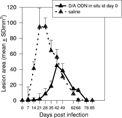
Oligodeoxynucleotides (ODN) containing CpG motifs mimic microbial DNA and are recognized by toll-like receptor 9 on immune cells. The resulting response limits the early spread of infectious organisms and promotes the development of adaptive immunity. In this regard, CpG ODN show promise as immunoprotective agents and as vaccine adjuvants. Previous studies of nonhuman primates showed that administration of CpG ODN type D (also known as type A) at the site of infection 3 days before and after a challenge with Leishmania major enhanced host resistance and reduced the lesion's severity. In this study, we show that systemic administration of D/A ODN limits the size of lesions following an intradermal infection with L. major. Importantly, the reduced morbidity was not associated with a reduction in long-term immunity, as such treated macaques were still protected following a secondary challenge. Finally, administration of D/A ODN to macaques that had established cutaneous lesions reduced the severity of the lesions, suggesting a potential role for CpG ODN in L. major treatment. Together, these findings support the development of clinical studies to assess the use of CpG ODN types D/A as immunoprotective and therapeutic agents.


Figures

FIG. 1.
FIG. 1.
Cutaneous lesions in macaques infected with L. major and treated with CpG ODN type D/A. Rhesus macaques were challenged with L. major (107 metacyclic promastigotes) i.d. on day zero. Three days before and after the challenge, the monkeys (n = 6/group) were treated with CpG ODN type D/A in situ (500 μg/animal i.d.) or systemically (500 μg/kg s.c. or i.m.). Untreated monkeys served as controls (n = 6). The average size of the lesions on the forehead is shown as the mean of the log normalized area [calculated as (mean diameter/2)2 × π]. Note that the lesions from macaques treated with D/A ODN peaked earlier and were smaller (P < 0.05) than those in untreated macaques. Differences among curves were tested by repeated-measures ANOVA using the Proc Mixed procedure. SD, standard deviation.
FIG. 2.
FIG. 2.
IFN-γ-secreting PBMC from macaques infected with L. major. Rhesus macaques were challenged with L. major and left untreated or treated with D/A ODN i.d. in situ (500 μg/dose), s.c., or i.m. (500 μg/kg/dose) 3 days before and after the challenge. The number of PBMC secreting IFN-γ upon in vitro stimulation with SLA was assessed by ELISPOT assay 3 weeks after challenge (a) or 4 months after the lesions were resolved (b). Shown are means plus standard errors of the mean for three to six macaques per group. Statistical differences were assessed by one-way ANOVA among the groups included in each key. NS, not significant.
FIG. 3.
FIG. 3.
Cutaneous lesions in macaques rechallenged with L. major. Eight months after the primary challenge (Fig. 1), macaques were rechallenged with L. major (LM; 2 × 10⁶ metacyclic promastigotes) i.d. on the contralateral forehead. Naïve macaques (n = 6) served as controls. The average size of the lesions on the forehead is shown as the mean of the log normalized area. Note that macaques that had been infected previously with L. major had reduced severity of lesions, regardless of treatment during the first challenge (P < 0.01). Differences among curves were tested by repeated-measures ANOVA using the Proc Mixed procedure from the Statistical Analysis System. SD, standard deviation.
FIG. 4.
FIG. 4.
IFN-γ-secreting PBMC from macaques rechallenged with L. major and stimulated in vitro with SLA. PBMC from rhesus macaques that had been infected with L. major and treated with D/A ODN and then rechallenged with 2 × 10⁶ L. major promastigotes were collected and tested for in vitro secretion of IFN-γ in response to SLA. PBMC from naïve infected animals served as controls. Shown are means plus standard errors of the mean for three to six macaques per group. Statistical significance was assessed by one-way ANOVA and Bonferroni's postcomparison procedure. NS, not significant.
FIG. 5.
FIG. 5.
Local parasite loads in monkeys after rechallenge with L. major. Rhesus macaques that had been challenged with L. major and left untreated or treated with D/A ODN i.d. in situ (500 μg), s.c., or i.m. (500 μg/kg) 3 days before and after the challenge were rechallenged with live parasites. Twenty days after rechallenge, the skin lesions were biopsied and the parasite loads were assessed. Shown are the parasite loads of individual monkeys. The biopsy procedure and estimation of parasite numbers were as described in Materials and Methods. Statistical differences among groups (P = 0.06) were tested using nonparametric Kruskal-Wallis one-way analysis of variance on ranks. ns, not significant.
FIG. 6.
FIG. 6.
Transient protection from lesions in macaques treated with D/A ODN at the time of infection. A group of rhesus macaques (n = 4) was treated with CpG ODN i.d. in situ (500 g/macaque) immediately following the infectious challenge (2 × 10⁶ metacyclic promastigotes). The area of the lesion developed was measured weekly. Note that the development of the lesions was delayed and reduced compared with macaques infected at the same time but left untreated (n = 6). SD, standard deviation.
FIG. 7.
FIG. 7.
Treatment of established L. major cutaneous lesions with D/A ODN. (a) Rhesus macaques infected with L. major (107 metacyclic promastigotes) i.d. on the forehead were either left untreated (control; n = 6) or treated (n = 6) with CpG ODN i.d. at the site of the lesion (500 μg/animal) 10 days after infection. (b) Rhesus macaques were infected i.d. with L. major (2 × 10⁶ metacyclic promastigotes) and either were left untreated (n = 6) or received CpG ODN type D/A s.c. in the interscapulary space (0.5 mg/kg; n = 4) on day 14. The means and standard deviations (SD) of the log normalized areas of the lesions are shown.

References

    1. Amaral, V. F., V. A. O. Ransatto, F. Conceicao-Solva, E. Molinaro, V. Ferreira, S. G. Coutinho, D. McMahon-Pratt, and G. Grimaldi. 1996. Leishmania amazonensis: the Asian rhesus macaques (macaca mulata) as an experimental model for the study of cutaneous leishmaniasis. Exp. Parasitol. 82:34-44. - PubMed
    1. Amaral, V. F., A. Teva, R. Porrozzi, A. J. Silva, M. S. Pereira, M. P. Oliveira-Neto, and G. Grimaldi, Jr. 2001. Leishmania major-infected rhesus macaques (Macaca mulatta) develop varying levels of resistance against homologous re-infections. Mem. Inst. Oswaldo Cruz 96:795-804. - PubMed
    1. Bauer, M., K. Heeg, H. Wagner, and G. B. Lipford. 1999. DNA activates human immune cells through a CpG sequence-dependent manner. Immunology 97:699-705. - PMC - PubMed
    1. Campos-Neto, A., R. Porrozzi, K. Greeson, R. N. Coler, J. R. Webb, Y. A. Seiky, S. G. Reed, and G. Grimaldi, Jr. 2001. Protection against cutaneous leishmaniasis induced by recombinant antigens in murine and nonhuman primate models of the human disease. Infect. Immun. 69:4103-4108. - PMC - PubMed
    1. Deng, J. C., T. A. Moore, M. W. Newstead, X. Zeng, A. M. Krieg, and T. J. Standiford. 2004. CpG oligodeoxynucleotides stimulate protective innate immunity against pulmonary Klebsiella infection. J. Immunol. 173:5148-5155. - PMC - PubMed

MeSH terms

LinkOut - more resources