. 2005 Aug;11(8):1317-28.
doi: 10.1261/rna.2520405.

The role of ribosome recycling factor in dissociation of 70S ribosomes into subunits

Affiliations

The role of ribosome recycling factor in dissociation of 70S ribosomes into subunits

Go Hirokawa et al. RNA. 2005 Aug.

Abstract

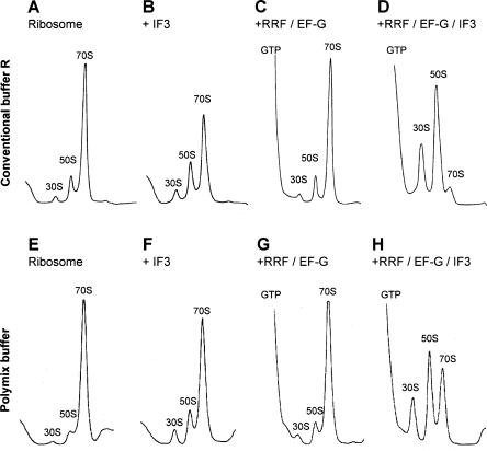
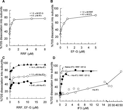
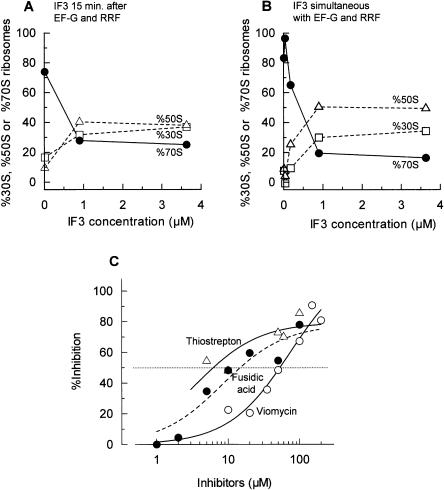
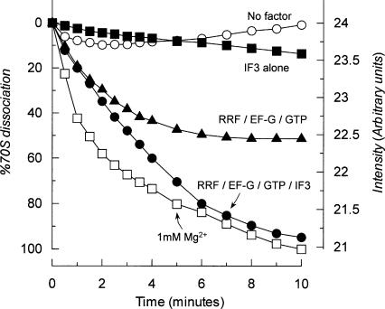
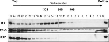
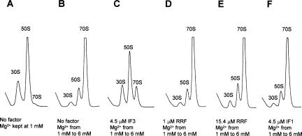
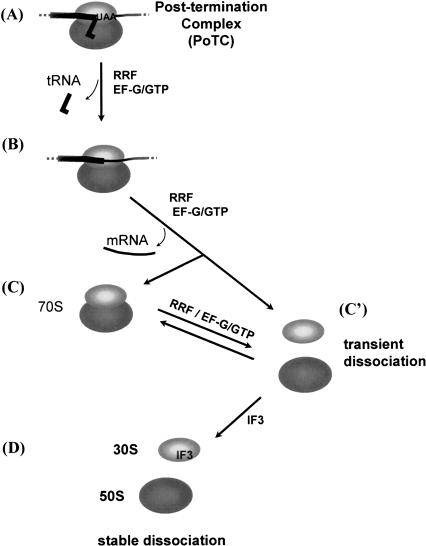
Protein synthesis is initiated on ribosomal subunits. However, it is not known how 70S ribosomes are dissociated into small and large subunits. Here we show that 70S ribosomes, as well as the model post-termination complexes, are dissociated into stable subunits by cooperative action of three translation factors: ribosome recycling factor (RRF), elongation factor G (EF-G), and initiation factor 3 (IF3). The subunit dissociation is stable enough to be detected by conventional sucrose density gradient centrifugation (SDGC). GTP, but not nonhydrolyzable GTP analog, is essential in this process. We found that RRF and EF-G alone transiently dissociate 70S ribosomes. However, the transient dissociation cannot be detected by SDGC. IF3 stabilizes the dissociation by binding to the transiently formed 30S subunits, preventing re-association back to 70S ribosomes. The three-factor-dependent stable dissociation of ribosomes into subunits completes the ribosome cycle and the resulting subunits are ready for the next round of translation.


Figures

FIGURE 1.
FIGURE 1.
In vivo concentrations of RRF, EF-G, and IF3 dissociate 70S ribosomes into subunits almost completely. W-ribosomes (0.07 μM) were incubated alone (A,E), with 4.5 μM IF3 (B,F), with 20 μM RRF, 20 μM EF-G, and 0.36 mM GTP (C,G), and with 20 μM RRF, 20 μM EF-G, 0.36 mM GTP, and 4.5 μM IF3 (D,H) at 30°C for 15 min in conventional buffer R (AD) or in the polymix buffer (EH). Sedimentation behavior of ribosomes was analyzed as described in Materials and Methods. The sedimentation is from left to right. The peaks of GTP, 30S, 50S, and 70S ribosome are indicated.
FIGURE 2.
FIGURE 2.
Dose-response curve of factors for the dissociation of 70S ribosomes into subunits. W-ribosomes (0.07 μM) were incubated with factors at 30°C in conventional buffer R. Sedimentation of ribosomes was analyzed as in Figure 1, and the amounts of 70S ribosomes converted into subunits are expressed as percentages. (A) RRF dose-response curve with 1 μM EF-G, 4.5 μM His-IF3, and 0.36 mM GTP. (B) EF-G dose-response curve with 1 μM RRF, 4.5 μM His-IF3, and 0.36 mM GTP. (C) RRF/EF-G dose-response curve. (•) 0.45 μM His-IF3, various amounts of RRF, EF-G, and 0.36 mM GTP. (○) As above except for 1 μM His-IF3. (▴) 14.4 μM His-IF3. (D) IF3 dose-response curve. ○, His-IF3; •, His-IF3, 5 μM RRF, 5 μM EF-G and 0.36 mM GTP; ▵ and dotted line, as above except that native IF3 was used instead of His-IF3.
FIGURE 3.
FIGURE 3.
Subunits dissociation from the model post-termination complexes by three factors. (A,B) IF3 dose-response curve. (A) Polysomes (0.6 A260 units) were incubated with puromycin (50 μM), RRF (1 μM), EF-G (1 μM), and GTP (0.36 mM) at 30°C for 10 min in conventional buffer R. Then, IF3 (various amounts) were added and further incubated at 30°C for 5 min. (B) Polysomes (0.6 A260 units) were incubated with puromycin (50 μM), RRF (1 μM), EF-G (1 μM), GTP (0.36 mM), and IF3 (various amounts) at 30°C for 15 min in conventional buffer R. Sedimentation behavior of ribosomes was analyzed as in Figure 1. The percentages of 30S subunits (□, dotted line), 50S subunits (▵, dotted line), and 70S ribosomes (•, solid line) are plotted against added IF3 concentrations. (C) Effects of EF-G inhibitors on the subunit dissociation from the model PoTC by RRF (1 μM), EF-G (1 μM), GTP (0.36 mM), and IF3 (4.5 μM) in conventional buffer R were examined as in B. ○ and solid line, viomycin; ▵ and solid line, thiostrepton; • and dotted line, fusidic acid. Percent inhibition of the increase of subunits was calculated and is plotted against the concentrations of inhibitors.
FIGURE 4.
FIGURE 4.
70S ribosomes are transiently dissociated by RRF and EF-G alone. (•) w-ribosomes (0.16 μM) were incubated with RRF (1 μM), EF-G (1 μM), GTP (0.36 mM), and IF3 (4.5 μM) at 30°C in buffer U, and the dissociation was observed following the decrease of the light scattering as described in Materials and Methods. (▴) With RRF (1 μM), EF-G (1 μM), and GTP (0.36 mM). (▪) With IF3 (4.5 μM) alone; ○ with no factors. (□) W-ribosomes (0.16 μM) were dissociated by placing into buffer S (1 mM Mg2+). The data are calculated as percentages of 70S ribosomes dissociated using the dissociation at 1 mM Mg2+ as 100%.
FIGURE 5.
FIGURE 5.
IF3, not RRF/EF-G, stays on subunits after the subunit formation by three factors. W-ribosomes (0.07 μM) were dissociated into subunits by 1 μM RRF, 1 μM EF-G, 0.36 mM GTP, and 4.5 μM IF3 in conventional buffer R, then sedimented through a 15%–30% sucrose gradient. Fractions were taken from the bottom of the sucrose gradient (10 drops per fraction, 24 fractions), and IF3, EF-G, and RRF were detected by Western blotting. Positions of 30S, 50S, and 70S are indicated. St (standard), purified IF3, EF-G, and RRF as controls.
FIGURE 6.
FIGURE 6.
RRF does not have anti-association activity. (A,B) W-ribosomes (0.07 μM) were incubated alone, (C) with 4.5 μM IF3, (D) with 1 μM RRF, (E) with 15.4 μM RRF, and (F) with 4.5 μM IF1, at 30°C for 5 min in buffer S (containing 1 mM MgSO4). Then, the MgSO4 concentration of BF, but not of A, was raised to 6 mM and further incubated at 30°C for 10 min. Ribosomes were sedimented through a 15%–30% sucrose density gradient in buffer S (A) or buffer T (containing 6 mM MgSO4; BF) and analyzed as in Figure 1. The sedimentation is from left to right. The peaks of 30S, 50S, and 70S ribosome are indicated.
FIGURE 7.
FIGURE 7.
Model of ribosomal subunit formation from a post-termination complex. (A) Post-termination complex with bound tRNA remained after releasing the nascent peptide. (B) Complex without tRNA due to the action of EF-G and RRF (Hirashima and Kaji 1973; Hirokawa et al. 2002). (C) 70S ribosome is free of mRNA and tRNA; (C′) transiently dissociated subunits by RRF and EF-G without IF3. (D) Stable subunits with IF3.

References

    1. Agrawal, R.K. and Burma, D.P. 1996. Sites of ribosomal RNAs involved in the subunit association of tight and loose couple ribosomes. J. Biol. Chem. 271: 21285–21291. - PubMed
    1. Agrawal, R.K., Sharma, M.R., Kiel, M.C., Hirokawa, G., Booth, T.M., Spahn, C.M.T., Grassucci, R.A., Kaji, A., and Frank, J. 2004. Visualization of ribosome-recycling factor on the Escherichia coli 70S ribosome: Functional implications. Proc. Natl. Acad. Sci. 101: 8900–8905. - PMC - PubMed
    1. Andersen, L.D., Moreno, J.M.D., Clark, B.F.C., Mortensen, K.K., and Sperling-Petersen, H.U. 1999. Immunochemical determination of cellular content of translation release factor RF4 in Escherichia coli. IUBMB Life 48: 283–286. - PubMed
    1. Bodley, J.W., Zieve, F.J., Lin, L., and Zieve, S.T. 1970. Studies on translocation III. Conditions necessary for the formation and detection of a stable ribosome-G factor-guanosine diphosphate complex in the presence of fusidic acid. J. Biol. Chem. 245: 5656–5661. - PubMed
    1. Caldas, T., Binet, E., Bouloc, P., and Richarme, G. 2000. Translational defects of Escherichia coli mutants deficient in the Um(2552) 23S ribosomal RNA methyltransferase RrmJ/FTSJ. Biochem. Biophys. Res. Commun. 271: 714–718. - PubMed

Publication types

MeSH terms

Substances

LinkOut - more resources