. 2005 Aug 1;202(3):405-13.
doi: 10.1084/jem.20050118. Epub 2005 Jul 25.

Cellular FLICE-inhibitory protein is required for T cell survival and cycling

Affiliations

Cellular FLICE-inhibitory protein is required for T cell survival and cycling

Hien Chau et al. J Exp Med. .

Abstract

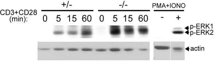
Fas-associated death domain (FADD) and caspase-8 are key signal transducers for death receptor-induced apoptosis, whereas cellular FLICE-inhibitory protein (cFLIP) antagonizes this process. Interestingly, FADD and caspase-8 also play a role in T cell development and T cell receptor (TCR)-mediated proliferative responses. To investigate the underlying mechanism, we generated cFLIP-deficient T cells by reconstituting Rag-/- blastocysts with cFLIP-deficient embryonic stem cells. These Rag chimeric mutant mice (rcFLIP-/-) had severely reduced numbers of T cells in the thymus, lymph nodes, and spleen, although mature T lymphocytes did develop. Similar to FADD- or caspase-8-deficient cells, rcFLIP-/- T cells were impaired in proliferation in response to TCR stimulation. Further investigation revealed that cFLIP is required for T cell survival, as well as T cell cycling in response to TCR stimulation. Interestingly, some signaling pathways from the TCR complex appeared competent, as CD3 plus CD28 cross-linking was capable of activating the ERK pathway in rcFLIP-/- T cells. We demonstrate an essential role for cFLIP in T cell function.


Figures

Figure 1.
Figure 1.
Expression of DED proteins in wild-type and FADD-DN T cells. Purified T cells isolated from FADD-DN transgenic mice and wild-type littermates were left untreated (0 h) or stimulated with 1 μg/ml anti-CD3 plus 5 μg/ml anti-CD28 antibodies for the time points indicated. Protein levels of cFLIP (long and short forms), FADD (full length and FADD-DN transgene), caspase-8, c-Myc, and actin were determined as described in Materials and methods. Arrows indicate the bands corresponding to the proteins of interest.
Figure 2.
Figure 2.
Generation of cFLIP-deficient T cells. (A) The targeting construct. Homologous recombination replaced a part of cFLIP exon 1, including the ATG start codon, with the neomycin resistance cassette on one of the wild-type alleles, resulting in a heterozygous ES cell line. A second targeting construct containing a hygromycin resistance cassette replaced the other allele, resulting in a cFLIP-null ES cell line. Primers used for PCR screening are indicated by arrows. (B) Absence of cFLIP expression confirmed by Northern blot analysis. 20 μg of total RNA was isolated from homozygous wild-type (+/+), homozygous mutant (−/−), and heterozygous (+/−) ES cell clones and resolved on a formaldehyde-denaturing agarose gel. RNA from a wild-type embryonic fibroblast cell line (EF) was also used as a control. The RNA was transferred to nitrocellulose membrane and probed with a full-length cFLIP cDNA probe (top) and a GADPH probe (bottom). (C) Expression of CD4 and CD8 in rcFLIP / peripheral T cells. Pooled lymph node cells from wild-type and rcFLIP / mice were stained for the expression of T cell–specific surface markers, CD4 and CD8, and analyzed by flow cytometry. The percentage of gated viable cells in each quadrant of the dot plots is shown. (D) Abnormal T cell development in rcFLIP / chimera. Representative flow cytometric analyses of thymi from rcFLIP / , rcFLIP + / , wild-type 129J, and Rag / mice are shown; the latter two were used as positive and negative controls, respectively. Thymocytes were stained using antibodies against CD4 and CD8 and analyzed by flow cytometry. The percentages of gated viable cells for each quadrant of the dot plots are shown. (E) Abnormal early thymocyte development in rcFLIP / chimera. Lineage-negative (CD4CD8) and Ly9.1-positive thymocytes from rcFLIP / and control mice (+/−) were gated and analyzed for the expression of CD25 and CD44. The percentage of gated cells in each quadrant is shown.
Figure 3.
Figure 3.
Defective proliferation of rcFLIP / T cells in response to TCR stimulation. (A) Defective proliferation of T cells lacking cFLIP in response to TCR stimulation. Purified T cells from spleens of rcFLIP + / (gray bars) and rcFLIP / (black bars) mice were stimulated with medium alone (untx), anti-CD28 alone, anti-CD3 (0.1 or 1 μg/ml) without or with 1 μg/ml anti-CD28, or 0.7 ng/ml IL-2. T cell proliferation was assessed by [3H]thymidine incorporation, and results shown were the mean counts per minute (cpm) ± SE from triplicate samples. (B) Abnormal cell division in CFSE-labeled rcFLIP / T cells. Purified CD4+ T cells from rcFLIP + / and rcFLIP / mice were labeled with CFSE and cultured with or without 1 μg/ml of plate-bound anti-CD3 antibody for 24, 48, or 72 h. Histograms show the CFSE fluorescence profile of viable, CFSE-labeled, CD4-positive T cells. The numbers above the peaks in each histogram denote the number of cell divisions the T cells have undergone; undivided T cells reside in the rightmost peak (denoted by 0). (C) Normal induction of early activation marker CD69. CD69 expression was measured by flow cytometry on rcFLIP / and control cells (+/−) treated with medium alone (untx), 1 μg/ml anti-CD3, or anti-CD3 plus 1 μg/ml anti-CD28 for 12 h. The numbers within each histogram represent the percentages of gated cells. (D) Normal expression of T cell activation marker CD25. CD25 (IL-2Rα) expression was measured by flow cytometry on rcFLIP / and control cells (+/−) similarly treated as in C for 24 h (top). The number within each histogram represents the percentage of gated cells. CD25 expression was assessed of cells within the 7-AAD–negative gate (outlined area), which is indicative of viable cells (bottom). The number above each dot plot represents the percentage of gated 7-AAD–negative cells.
Figure 4.
Figure 4.
Survival defect of rcFLIP / T cells. (A) Enhanced spontaneous and TCR-induced cell death in T cells lacking cFLIP. Purified T cells from rcFLIP + / and rcFLIP / mice were stimulated with medium alone (untx) or 1 μg/ml of insoluble anti-CD3 antibody for 48 h. Cells were then stained with anti-Thy1.2, Annexin V, and 7-AAD and examined by flow cytometry. Analysis was performed on Thy1.2-positive cells. The percentage of gated cells in each quadrant is shown. (B) Cell viability histogram of purified T cells from rcFLIP + / (gray bars) and rcFLIP / (black bars) mice stimulated with medium alone (untx), 1 μg/ml anti-CD3, or anti-CD3 plus 5 μg/ml anti-CD28 for 24 and 48 h. Cell viability was assessed by flow cytometric analysis of 7-AAD–negative cells, and the results shown were the average percentage of cell viability ± SD for duplicate samples. (C) The role of the Fas signal on rcFLIP / T cell proliferation. Purified T cells from rcFLIP + / and rcFLIP / mice were treated with medium alone (untx), 1 μg/ml of insoluble anti-CD3 antibody without or with 1 μg/ml anti-CD28 antibody in the absence or presence of 10 μg/ml of neutralizing anti-FasL antibody for 24 h. T cell proliferation was assessed by [3H]thymidine incorporation, and results shown are cpm ± SE from triplicate samples.
Figure 5.
Figure 5.
Impaired rcFLIP / T lymphocyte proliferation in response to TCR activation. (A, top) Viable cell counts by trypan blue exclusion from duplicate wells made of rcFLIP + / and rcFLIP / T cells after 48 h of culture without or with 1 μg/ml anti-CD3 treatment. Approximately twice as many rcFLIP / cells were seeded in each well as compared with rcFLIP + / cells. (bottom) Defective thymidine incorporation of rcFLIP / T cells in response to TCR stimulation. rcFLIP + / (white bars) and rcFLIP / (black and gray bars) T cells were cultured without or with 1 μg/ml anti-CD3 treatment as described in A (top), and proliferation was measured by [3H]thymidine incorporation with results shown as mean cpm ± SE. (B) Impairment of cell cycle entry in rcFLIP / T cells. Purified T cells from rcFLIP + / and rcFLIP / mice were either untreated (untx) or treated with 1 μg/ml of plate-bound anti-CD3 antibody with or without 1 μg/ml anti-CD28 antibody. Cells were fixed and stained with FITC-labeled anti-BrdU antibody and 7-AAD. Cells in the different phases of the cell cycle (G0/G1, S, and G2/M) are indicated. The percentage of cells in the S and G2/M phases of the cell cycle is shown.
Figure 6.
Figure 6.
Competent TCR-induced ERK activation in rcFLIP / T cells. Competent activation of Erk in rcFLIP / T cells. Purified T cells from rcFLIP + / and rcFLIP / mice were untreated (0 min) or activated with 10 μg/ml each of soluble anti-CD3 and anti-CD28 antibodies for the indicated time points. PMA plus calcium ionophore (100 ng/ml each) stimulation (+) compared with no treatment (−) of purified T cells from wild-type (129J strain) mice was used as a positive control for Erk phosphorylation. Phosphorylated Erk was detected using phosphospecific antibodies by Western blotting. Actin was used as a loading control. Arrows indicate the bands corresponding to the proteins of interest.

References

    1. Chen, G., and D.V. Goeddel. 2002. TNF-R1 signaling: a beautiful pathway. Science. 296:1634–1635. - PubMed
    1. Tibbetts, M.D., L. Zheng, and M.J. Lenardo. 2003. The death effector domain protein family: regulators of cellular homeostasis. Nat. Immunol. 4:404–409. - PubMed
    1. Barnhart, B.C., J.C. Lee, E.C. Alappat, and M.E. Peter. 2003. The death effector domain protein family. Oncogene. 22:8634–8644. - PubMed
    1. Aggarwal, B.B. 2003. Signalling pathways of the TNF superfamily: a double-edged sword. Nat. Rev. Immunol. 3:745–756. - PubMed
    1. Thorburn, A. 2004. Death receptor-induced cell killing. Cell. Signal. 16:139–144. - PubMed

Publication types

MeSH terms