. 2005 Aug 2;102(31):11017-22.
doi: 10.1073/pnas.0504823102. Epub 2005 Jul 25.

HIV-1 Nef down-regulates the hemochromatosis protein HFE, manipulating cellular iron homeostasis

Affiliations

HIV-1 Nef down-regulates the hemochromatosis protein HFE, manipulating cellular iron homeostasis

Hal Drakesmith et al. Proc Natl Acad Sci U S A. .

Abstract

The multifunctional Nef protein of HIV-1 is important for the progression to AIDS. One action of Nef is to down-regulate surface MHC I molecules, helping infected cells to evade immunity. We found that Nef also down-regulates the macrophage-expressed MHC 1b protein HFE, which regulates iron homeostasis and is mutated in the iron-overloading disorder hemochromatosis. In model cell lines, Nef reroutes HFE to a perinuclear structure that overlaps the trans-Golgi network, causing a 90% reduction of surface HFE. This activity requires a Src-kinase-binding proline-rich domain of Nef and a conserved tyrosine-based motif in the cytoplasmic tail of HFE. HIV-1 infection of ex vivo macrophages similarly down-regulates naturally expressed surface HFE in a Nef-dependent manner. The effect of Nef expression on cellular iron was explored; iron and ferritin accumulation were increased in HIV-1-infected ex vivo macrophages expressing wild-type HFE, but this effect was lost with Nef-deleted HIV-1 or when infecting macrophages from hemochromatosis patients expressing mutated HFE. The iron accumulation in HIV-1-infected HFE-expressing macrophages was paralleled by an increase in cellular HIV-1-gag expression. We conclude that, through Nef and HFE, HIV-1 directly regulates cellular iron metabolism, possibly benefiting viral growth.


Figures

Fig. 1.
Fig. 1.
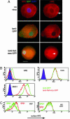
Down-regulation and redistribution of surface HFE and its ligand TfR by HIV-1 Nef in cell lines. (A) Alignment of amino acid sequence of the cytoplasmic portion of HLA-A2 and HFE. Conserved residues are shown in red, and the YxxA motif is underlined. (B) HeLa-HFE cells were transiently transfected with Nef-GFP, stained for surface HFE, TfR, MHC I, or epidermal growth factor receptor (EGFR), and analyzed by flow cytometry. Results show that, as Nef expression increases (along the x axis), surface levels of HFE, TfR, and, to a lesser extent, MHC I decrease (y axis), whereas EGFR expression is unchanged. (C) Aliquots of 293 cells stably expressing HFE or HFE mutants, as indicated, were transiently transfected with Nef-GFP, and Nef-positive and Nef-negative cells were assayed for surface HFE and TfR. The results show that Nef-mediated down-regulation of HFE and TfR depends on a cytoplasmic tyrosine 342Yof HFE and correct folding of HFE (the C282Y mutation misfolds and does not bind TfR). (D-I) HeLa-HFE cells were transiently transfected with Nef-GFP, permeabilized, and stained for HFE (E and F, red) and for TGN46 (reveals the TGN) (H and I, red). Nef (D, F, G, and I, green) locates mostly to a perinuclear structure overlapping the TGN (I, yellow; nuclei stained blue with DAPI). HFE (E and F, red) locates to the cell membrane and endosomes in untransfected cells (E, arrow and F Left) but is redistributed to a perinuclear intracellular region overlapping Nef in transfected cells (E, arrowheads and F, yellow). wt, wild type. (Scale bar, 10 μm.)
Fig. 2.
Fig. 2.
Down-regulation of endogenous surface HFE in monocytic THP-1 cells and primary macrophages by Nef and HIV. (A) Ex vivo macrophages were infected with recombinant lentivirus-based vectors encoding either GFP (lenti-GFP) or Nef-IRES-GFP (lenti-Nef-IRES-GFP) and stained for HFE (red). Images of representative cells show that surface HFE expression on membrane ruffles in uninfected cells is unaltered in cells infected with the control lenti-GFP virus (Top and Middle Right, arrows) but that HFE is redistributed to a perinuclear region in cells infected with lenti-Nef-IRES-GFP (Bottom Right, arrowhead). (Scale bar, 10 μm.) (B) The cell monocyte/macrophage cell line THP-1 normally expresses low levels of surface HFE that are unchanged by the control lenti-GFP virus, but, in cells infected with lenti-Nef-IRES-GFP, surface HFE is undetectable above the background. Nef does not alter surface CD29 (negative control) but MHC I levels are lowered (positive control). (C) Macrophages were infected for 4 days with M-tropic HIV-1, with (HIV) or without (HIVΔNef) the nef gene, stained for surface HFE (anti-α3 domain) and p18/p55 gag, and analyzed by FACS. HIV-infected gag-expressing cells (Left, green line) express less surface HFE than do uninfected cells from the same culture (Left, red line). HIVΔNef infection (Right) does not alter surface HFE. This finding was repeated on macrophages from multiple donors expressing wild-type HFE.
Fig. 3.
Fig. 3.
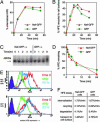
Mechanism of Nef-mediated HFE down-regulation. (A) Internalization of HFE. HeLa-HFE cells transfected with Nef-GFP or GFP for 2 days were labeled with anti-HFE antibody at 4°C and then washed and incubated for given times at 37°C to allow internalization. Remaining surface HFE was stained with anti-mouse Ig antibody, and cells were analyzed by FACS. The percentage of internalized HFE was calculated and plotted. (B) Recycling of HFE. Surface β2-microglobulin was stripped from cycloheximide-pretreated Nef-GFP or GFP-expressing HeLa-HFE cells, and the appearance of surface β2-microglobulin-associated HFE from intracellular compartments was assayed, over time, by using FACS. (C and D) Degradation of HFE over time. HeLa-HFE cells transfected with Nef-GFP or GFP were pulsed with [35S]methionine and chased with excess cold methionine for the indicated times. HFE was immunoprecipitated from cell lysates and analyzed by PAGE (C). Numbers above the gel indicate hours of chase after 35S-labeling. Densitometric analysis (D) of gels from several experiments similar to that shown in C allowed quantification of the percentage of remaining HFE. (E) Transport of HFE to the cell surface. Aliquots of HeLa-HFE cells were transfected for 15 h with Nef-GFP (Upper) or GFP (Lower). Cells were stained for surface HFE (red line; at this early time point, Nef has not yet down-regulated HFE), stripped of surface β2-microglobulin and stained again for HFE (green line) or stripped and cultured for 20 h in serum-free medium to allow transport to the cell surface of newly synthesized HFE/β2-microglobulin molecules and stained for HFE (blue line). In Nef-expressing cells, newly synthesized HFE reaching the cell surface is reduced. (F) Summary of A-E: rates of internalization and recycling over 30 min did not differ with expression of Nef. Nef marginally increased HFE degradation, and Nef reduced transport of HFE to the cell surface over 20 h by 3.2-fold.
Fig. 4.
Fig. 4.
Effects of mutations in Nef on Nef-mediated down-regulation of HFE, TfR, and MHC I. (A-D) HeLa-HFE cells were transfected with Nef-GFP constructs (x axis) encoding wild-type Nef (A), 62EEEE65/AAAA Nef (B), 20M/A Nef (C), and 72PxxP75/AxxA Nef (D) and stained for surface HFE (Top, y axis), TfR (Middle, y axis), and MHC I (Bottom, y axis). The results show that the ability of Nef to down-regulate HFE and TfR is retained by Nef variants lacking 62EEEE65 or 20M, but the 72PxxP75 motif is required for HFE/TfR down-regulation. All three motifs are needed for down-regulation of MHC I. (E) The role of other Nef motifs. HeLa-HFE cells were transfected with the Nef-GFP variants indicated and stained for surface MHC I, HFE, and TfR. The percentage of down-regulation, relative to down-regulation by wild-type Nef-GFP (100%), was calculated. Myristolation and the proline-rich domain are required for MHC I, HFE, and TfR down-regulation; four Nef motifs, needed for CD4 down-regulation, are dispensable.
Fig. 5.
Fig. 5.
The SH3 domain of dominant negative Hck blocks Nef-mediated down-regulation of HFE. (A) Wild-type Hck has SH3, SH2, and a catalytic domain. Dominant negative HckN lacks the catalytic domain. (B) HeLa-HFE cells transfected with Nef-GFP (green) have reduced surface HFE, compared with Nef-negative cells (red). (C) Cotransfection at 1:1 of Nef-GFP with HckN blocks the effect of Nef on HFE. (D and E) A W93F HckN variant with dysfunctional SH3 domains (D) does not block Nef activity, whereas the blocking effect of HckN is maintained when SH2-disrupted R151S Hck (E) is used. (F) CrkII (which has SH3 domains but does not bind Nef) does not affect HFE down-regulation by Nef. (G) Nef-GFP is normally located perinuclearly, overlapping the TGN, but coexpression of HckN (H) reroutes Nef-GFP to the cell periphery. In the presence of HckN, Nef-GFP no longer colocalizes with the TGN (I, nuclei stained blue with DAPI), and HFE resumes its endosomal localization (J), similar to that in untransfected cells. (Scale bars, 10 μm.)
Fig. 6.
Fig. 6.
The Nef-HFE interaction increases iron accumulation and HIV growth in monocytes/macrophages. (A) THP-1 cells were infected with lentiviral vectors encoding GFP or Nef-GFP and then permeabilized and analyzed for ferritin protein levels. Ex vivo macrophages homozygous for wild-type HFE or C282Y HFE were infected for 4 days with 50 ng/ml p24 HIV or Nef-deleted HIV and stained for p18/p55 gag and ferritin. The mean fluorescence intensity of 30,000-cell aliquots is given. Nef-GFP-expressing THP-1 cells contained double the ferritin of uninfected cells; GFP-infected cells have only a small increase in ferritin. HIV infection increases ferritin only in wild-type-expressing macrophages, and this effect is reduced in the absence of Nef. (B and C) THP-1 (B) and U937 (C), a monocytic cell line that does not express HFE, were transfected by nucleoporation with either Nef-GFP or GFP and incubated with 40 μg/ml [59Fe]transferrin for 14 h, and iron accumulation was measured. Nef-GFP increased intracellular iron in THP-1 but not in U937 cells. The mean accumulation was calculated, and the asterisk indicates significance, compared with GFP control (P < 0.001 by Student's t test). (D) Ex vivo macrophages homozygous for either wild-type or C282Y HFE were infected with wild-type HIV or Nef-deleted HIV and, after 2 days, incubated with 33 μg/ml [59Fe]transferrin for an additional 10 days. The cellular 59Fe was then determined. HIV increased macrophage iron accumulation in a Nef- and wild-type-HFE-dependent manner. Experiments shown in A-D are representative of several giving similar results. (E) Ex vivo macrophages homozygous for wild-type HFE (four donors) or C282Y HFE (three donors) were incubated with 50 ng/ml p24 of M-tropic HIV, with or without nef, for 4 days, stained for p18/p55 gag expression, and analyzed by FACS to calculate the percentage of p18/p55-positive cells in the cultures. The data show that Nef induces a 2- to 3-fold increase in gag-expressing cells in wild-type HFE-expressing macrophages, but this effect is reduced in C282Y-expressing macrophages and is not induced by Nef.

Similar articles

Cited by

References

    1. Deacon, N. J., Tsykin, A., Solomon, A., Smith, K., Ludford-Menting, M., Hooker, D. J., McPhee, D. A., Greenway, A. L., Ellett, A., Chatfield, C., et al. (1995) Science 270, 988-991. - PubMed
    1. She, H., Xiong, S., Lin, M., Zandi, E., Giulivi, C. & Tsukamoto, H. (2002) in Am. J. Physiol. 283, G719-G726. - PubMed
    1. Geyer, M., Fackler, O. T. & Peterlin, B. M. (2001) EMBO Rep. 2, 580-585. - PMC - PubMed
    1. Guy, B., Kieny, M. P., Riviere, Y., Le Peuch, C., Dott, K., Girard, M., Montagnier, L. & Lecocq, J. P. (1987) Nature 330, 266-269. - PubMed
    1. Aiken, C., Konner, J., Landau, N. R., Lenburg, M. E. & Trono, D. (1994) Cell 76, 853-864. - PubMed

Publication types

MeSH terms