. 2005 Aug 2;102(31):11076-81.
doi: 10.1073/pnas.0504266102. Epub 2005 Jul 25.

Iron and Pseudomonas aeruginosa biofilm formation

Affiliations

Iron and Pseudomonas aeruginosa biofilm formation

Ehud Banin et al. Proc Natl Acad Sci U S A. .

Abstract

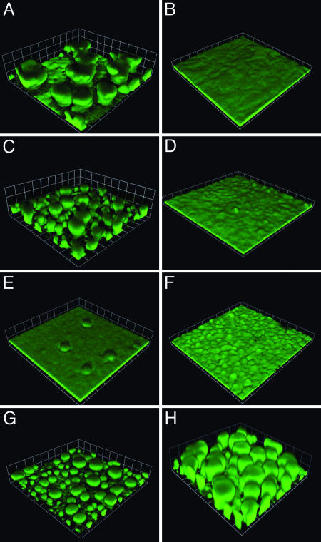
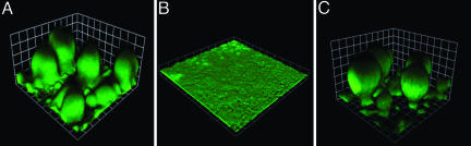
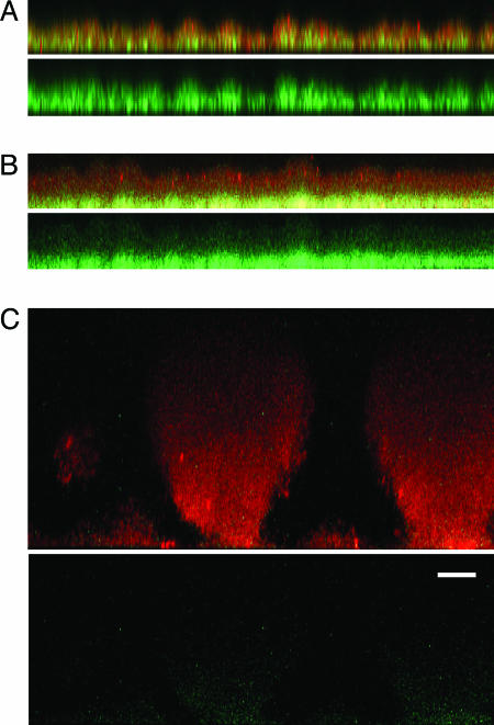
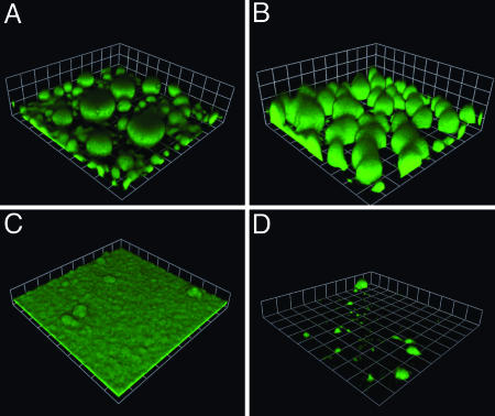
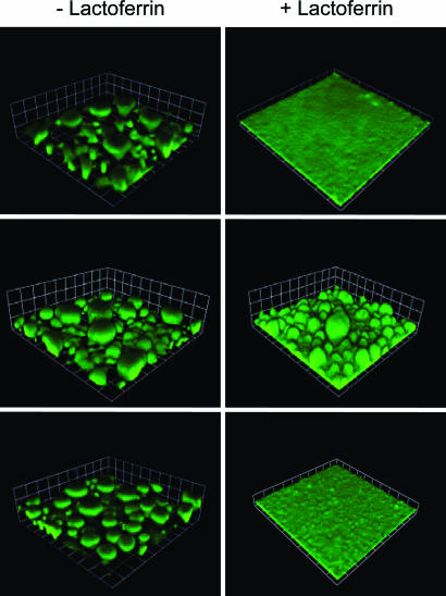
Iron serves as a signal in Pseudomonas aeruginosa biofilm development. We examined the influence of mutations in known and putative iron acquisition-signaling genes on biofilm morphology. In iron-sufficient medium, mutants that cannot obtain iron through the high-affinity pyoverdine iron acquisition system form thin biofilms similar to those formed by the parent under low iron conditions. If an iron source for a different iron acquisition system is provided to a pyoverdine mutant, normal biofilm development occurs. This enabled us to identify iron uptake gene clusters that likely serve in transport of ferric citrate and ferrioxamine. We suggest that the functional iron signal for P. aeruginosa biofilm development is active transport of chelated iron or the level of internal iron. If the signal is internal iron levels, then a factor likely to be involved in iron signaling is the cytoplasmic ferric uptake regulator protein, Fur, which controls expression of iron-responsive genes. In support of a Fur involvement, we found that with low iron a Fur mutant was able to organize into more mature biofilms than was the parent. The two known Fur-controlled small regulatory RNAs (PrrF1 and F2) do not appear to mediate iron control of biofilm development. This information establishes a mechanistic basis for iron control of P. aeruginosa biofilm formation.


Figures

Fig. 1.
Fig. 1.
Biofilm formation of P. aeruginosa pyoverdine and pyochelin mutants. The parent without (A) and with (B) lactoferrin (20 μg/ml), the pyochelin mutant PAO1pchA::TcR without (C) and with (D) lactoferrin (20 μg/ml), the pyoverdine synthesis mutant PAO1ΔpvdA (E), the pyoverdine ECF σ-factor PvdS mutant PAOΔpvdS without lactoferrin (F), the pyoverdine mutant carrying the pvdA expression vector pEB4 without lactoferrin (G), and the pyoverdine mutant grown in pyoverdine-conditioned medium without lactoferrin (H). The P. aeruginosa cells contained the gfp plasmid pMRP9-1. Images are from 6-day biofilms (the squares are 61 μm on a side).
Fig. 2.
Fig. 2.
Active iron transport is required for normal biofilm development in flow cells. (A) Biofilm of the pvdA mutant PAO1ΔpvdA grown with added ferric chloride (50 μM). Biofilm of a pyoverdine, pyochelin double mutant (EB107) grown with 100 μM ferric chloride (B) or 1.5 μM desferrioxamine (C) (similar results were obtained when 1 μM ferric dicitrate was added; data not shown). All strains contained the gfp plasmid pMRP9-1. Images represent 6-day biofilms (the squares are 30 μm on a side).
Fig. 3.
Fig. 3.
Expression of pvdS-gfp in biofilms of a pyoverdine, pyochelin double mutant (EB125) in the presence of FeCl3 or desferrioxamine. (A) Biofilms grown without FeCl3.(B) Biofilms grown with 100 μM FeCl3.(C) Biofilms grown with 1.5 μM desferrioxamine. Images represent 6-day biofilms counterstained with propidium iodide. (A-C Upper) A reconstructed side view of the combined red and green channels. Red shows the biofilm, and green shows regions where cells have expressed GFP. (A-C Lower)A side view of GFP expression alone. (Scale bar in Lower, 15 μm; all of the side images are the same magnification.)
Fig. 4.
Fig. 4.
Ferric citrate and ferrioxamine mediate development of mushroom-like structures in the pyoverdine synthesis mutant PAO1ΔpvdA. The pvdA mutant biofilm in medium supplemented with 1 μM ferric dicitrate (A) or 1.5 μM desferrioxamine (B). (C) A biofilm of the pvdA, PA3901 double mutant EB104 in medium containing 1 μM ferric dicitrate. (D) A biofilm of EB112 (the pyoverdine, PA2466, and PA0470 triple mutant) in medium with 1.5 μM desferrioxamine. All strains contained pMRP9-1. Images represent 6-day biofilms (the squares are 62 μm on a side).
Fig. 5.
Fig. 5.
Biofilm formation by Fur and PrrF mutants. (Top) Biofilms of the parent PAO1. (Middle) Biofilms of the Fur mutant PAO1furC6Tc. (Bottom) Biofilms of the PAO1ΔprrF1-F2 double mutant. Where indicated, biofilms were grown in the presence of 20 μg/ml lactoferrin. Strain PAO1 and PAO1ΔprrF1-F2 contained the gfp plasmid pMRP9-1. The Fur mutant was stained with propidium iodide (we could not introduce pMRP9-1 into the strain). Images in Left are of 6-day biofilms, and the squares are 60 μm on a side. Images in Right are also images of 6-day biofilms, but the squares are 30 μm on a side.

References

    1. Costerton, J. W., Stewart, P. S. & Greenberg, E. P. (1999) Science 284, 1318-1322. - PubMed
    1. Hall-Stoodley, L., Costerton, J. W. & Stoodley, P. (2004) Nat. Rev. Microbiol. 2, 95-108. - PubMed
    1. Lyczak, J. B., Cannon, C. L. & Pier, G. B. (2000) Microbes Infect. 2, 1051-1060. - PubMed
    1. Singh, P. K., Parsek, M. R., Greenberg, E. P. & Welsh, M. J. (2002) Nature 417, 552-555. - PubMed
    1. Singh, P. K. (2004) Biometals 17, 267-270. - PubMed

Publication types

MeSH terms