. 2005 Aug 2;102(31):10976-80.
doi: 10.1073/pnas.0505077102. Epub 2005 Jul 25.

The molecular structure of the Toll-like receptor 3 ligand-binding domain

Affiliations

The molecular structure of the Toll-like receptor 3 ligand-binding domain

Jessica K Bell et al. Proc Natl Acad Sci U S A. .

Abstract

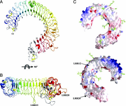
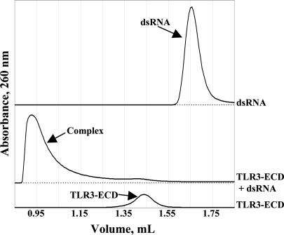
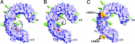
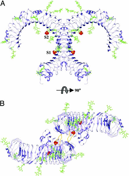
Innate immunity is the first line of defense against invading pathogens. Toll-like receptors (TLRs) act as sentinels of the innate immune system, sensing a variety of ligands from lipopolysaccharide to flagellin to dsRNA through their ligand-binding domain that is composed of leucine-rich repeats (LRRs). Ligand binding initiates a signaling cascade that leads to the up-regulation of inflammation mediators. In this study, we have expressed and crystallized the ectodomain (ECD) of human TLR3, which recognizes dsRNA, a molecular signature of viruses, and have determined the molecular structure to 2.4-A resolution. The overall horseshoe-shaped structure of the TLR3-ECD is formed by 23 repeating LRRs that are capped at each end by specialized non-LRR domains. The extensive beta-sheet on the molecule's concave surface forms a platform for several modifications, including insertions in the LRRs and 11 N-linked glycans. The TLR3-ECD structure indicates how LRR loops can establish distinct pathogen recognition receptors.


Figures

Fig. 1.
Fig. 1.
Overview of TLR3-ECD structure. (A) TLR3-ECD is shown in a ribbon diagram with the N- and C-terminal capping motifs colored in gray, the LRRs colored from blue to red beginning at LRR1, and the four disulfide bonds depicted by yellow ball-and-stick. (B) Side view colored as in A shows the two insertions at LRRs 12 and 20, respectively. (C) Electrostatic potential surface shows positive (blue) and negative (red) charges at neutral pH. The N-linked glycans are shown as green ball-and-stick. One surface is covered in glycan (Upper), whereas the opposite face (Lower) is devoid of glycan with a pocket of basic residues bounded by the two LRRs containing insertions 12 and 20, respectively.
Fig. 2.
Fig. 2.
dsRNA binding to TLR3-ECD. TLR3-ECD and dsRNA form a large, aggregated complex, as observed by gel filtration.
Fig. 3.
Fig. 3.
Proposed modes of ligand recognition for TLR3-ECD. Surface rendering of the TLR3-ECD shows the protein core in purple, glycan in green, sulfate ions in red, and LRR12 and -20 insertions in orange. A 19-mer of dsRNA shown in gray ribbons was docked into two potential binding sites (A and B) based on the location of sulfate ions that may mimic nucleic phosphate backbone interactions. (C) The unglycosylated surface of the TLR3-ECD offers a third potential docking site with a shallow groove bounded by the insertions in LRRs 12 and 20.
Fig. 4.
Fig. 4.
Crystal packing of TLR3-ECD. Ribbon diagram of the TLR3-ECD symmetry-related dimer found in the crystal lattice. N-glycosylation is shown in green ball-and-stick, and the two sulfate ions are shown in yellow and red space-filling (S1 and S2). The side view (A) shows the predominance of glycan decorating the faces of the molecule. Looking down the crystallographic twofold between the molecules (B), a surface near the C terminus that is devoid of glycan allows for close contact between molecules. The interface contains four hydrogen bonds, one of which (Asp-575) is contributed by the insertion in LRR20 (shown in orange).

References

    1. Takeda, K., Kaisho, T. & Akira, S. (2003) Annu. Rev. Immunol. 21, 335-376. - PubMed
    1. Takeda, K. & Akira, S. (2005) Int. Immunol. 17, 1-14. - PubMed
    1. Lemaitre, B., Nicolas, E., Michaut, L., Reichhart, J. M. & Hoffmann, J. A. (1996) Cell 86, 973-983. - PubMed
    1. Beutler, B. & Rehli, M. (2002) Curr. Top. Microbiol. Immunol. 270, 1-21. - PubMed
    1. Akira, S. & Takeda, K. (2004) Nat. Rev. Immunol. 4, 499-511. - PubMed

Publication types

MeSH terms

Associated data