Galanin receptors in the rat gastrointestinal tract
- PMID: 16044511
- PMCID: PMC3846504
- DOI: 10.1016/j.npep.2004.12.023
Galanin receptors in the rat gastrointestinal tract
Abstract
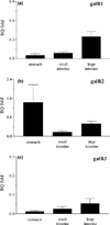
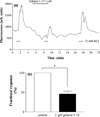
Galanin functions are mediated by three distinct G-protein-coupled receptors, galanin receptor 1 (GalR1), GalR2 and GalR3, which activate different intracellular signaling pathways. Here, we quantified mRNA levels of GalR1, GalR2 and GalR3 in the gastrointestinal tract using real time RT-PCR. GalR1 and GalR2 mRNAs were detected in all segments with the highest levels in the large intestine and stomach, respectively. GalR3 mRNA levels were quite low and mostly confined to the colon. We also investigated the effect of galanin 1-16, which has high affinity for GalR1 and GalR2 and low affinity for GalR3 on depolarization-evoked Ca2+ increases in rat cultured myenteric neurons using Ca2+-imaging. Intracellular Ca2+ changes in myenteric neurons were monitored using the Ca2+ sensitive dye, fluo-4, and confocal microscopy. Galanin 1-16 (1 microM) markedly inhibited the K+-evoked Ca2+ increases in myenteric neurons. In summary, the differential distribution of GalRs supports the hypothesis that the complex effects of galanin in the gastrointestinal tract result from the activation of multiple receptor subtypes. Furthermore, this study confirms the presence of functional GalRs and suggests that galanin modulates transmitter release from myenteric neurons through inhibition of voltage-dependent calcium channels involving a G(i/o)-coupled GalR.
Figures


References
-
- Branchek TA, Smith KE, Walker MW. Molecular biology and pharmacology of galanin receptors. Ann. N.Y. Acad. Sci. 1998;863:94–107. - PubMed
-
- Branchek TA, Smith KE, Gerald C, Walker MW. Galanin receptor subtypes. Trends Pharmacol. Sci. 2000;21:109–117. - PubMed
-
- Fontaine J, Lebrun P. Galanin: Ca2+-dependent contractile effects on the isolated mouse distal colon. Eur. J. Pharmacol. 1989;164:583–586. - PubMed
Publication types
MeSH terms
Substances
Grants and funding
LinkOut - more resources
Full Text Sources
Molecular Biology Databases
Miscellaneous

