

Putative DNA quadruplex formation within the human c-kit oncogene
- PMID: 16045346
- PMCID: PMC2195896
- DOI: 10.1021/ja050823u
Putative DNA quadruplex formation within the human c-kit oncogene
Abstract
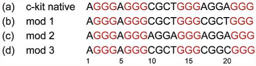
The DNA sequence, d(AGGGAGGGCGCTGGGAGGAGGG), occurs within the promoter region of the c-kit oncogene. We show here, using a combination of NMR, circular dichroism, and melting temperature measurements, that this sequence forms a four-stranded quadruplex structure under physiological conditions. Variations in the sequences that intervene between the guanine tracts have been examined, and surprisingly, none of these modified sequences forms a quadruplex arrangement under these conditions. This suggests that the occurrence of quadruplex-forming sequences within the human and other genomes is less than was hitherto expected. The c-kit quadruplex may be a new target for therapeutic intervention in cancers where there is elevated expression of the c-kit gene.
Figures


Similar articles
-
A conserved quadruplex motif located in a transcription activation site of the human c-kit oncogene.Biochemistry. 2006 Jun 27;45(25):7854-60. doi: 10.1021/bi0601510. Biochemistry. 2006. PMID: 16784237 Free PMC article.
-
Solution structures of all parallel-stranded monomeric and dimeric G-quadruplex scaffolds of the human c-kit2 promoter.Nucleic Acids Res. 2010 Oct;38(19):6757-73. doi: 10.1093/nar/gkq558. Epub 2010 Jun 21. Nucleic Acids Res. 2010. PMID: 20566478 Free PMC article.
-
Polymorphism and resolution of oncogene promoter quadruplex-forming sequences.Org Biomol Chem. 2011 Oct 26;9(22):7633-7. doi: 10.1039/c1ob05891f. Epub 2011 Sep 21. Org Biomol Chem. 2011. PMID: 21938285 Free PMC article.
-
Structures, folding patterns, and functions of intramolecular DNA G-quadruplexes found in eukaryotic promoter regions.Biochimie. 2008 Aug;90(8):1149-71. doi: 10.1016/j.biochi.2008.02.020. Epub 2008 Feb 29. Biochimie. 2008. PMID: 18355457 Free PMC article. Review.
-
Making sense of G-quadruplex and i-motif functions in oncogene promoters.FEBS J. 2010 Sep;277(17):3459-69. doi: 10.1111/j.1742-4658.2010.07759.x. Epub 2010 Jul 29. FEBS J. 2010. PMID: 20670278 Free PMC article. Review.
Cited by
-
G-Quadruplex Structures in Bacteria: Biological Relevance and Potential as an Antimicrobial Target.J Bacteriol. 2021 Jun 8;203(13):e0057720. doi: 10.1128/JB.00577-20. Epub 2021 Jun 8. J Bacteriol. 2021. PMID: 33649149 Free PMC article. Review.
-
The interaction of telomeric DNA and C-myc22 G-quadruplex with 11 natural alkaloids.Nucleic Acid Ther. 2012 Apr;22(2):127-36. doi: 10.1089/nat.2012.0342. Nucleic Acid Ther. 2012. PMID: 22480315 Free PMC article.
-
Classification of g-quadruplex DNA on the basis of the quadruplex twist angle and planarity of g-quartets.Acta Naturae. 2010 Oct;2(4):72-81. Acta Naturae. 2010. PMID: 22649667 Free PMC article.
-
Molecular dynamics and principal components of potassium binding with human telomeric intra-molecular G-quadruplex.Protein Cell. 2015 Jun;6(6):423-33. doi: 10.1007/s13238-015-0155-3. Epub 2015 Apr 18. Protein Cell. 2015. PMID: 25894091 Free PMC article.
-
Quadruplex DNA: sequence, topology and structure.Nucleic Acids Res. 2006;34(19):5402-15. doi: 10.1093/nar/gkl655. Epub 2006 Sep 29. Nucleic Acids Res. 2006. PMID: 17012276 Free PMC article. Review.
References
Publication types
MeSH terms
Substances
Grants and funding
LinkOut - more resources
Full Text Sources
Other Literature Sources
